Deep Veins

MYO CORE

Vertebral Venous Plexuses

The vertebral venous plexus is an extensive valveless longitudinal network comprising the internal and external vertebral venous plexuses interconnected by the intervertebral veins. Extending from the cranial cavity to the sacrum, it forms the principal collateral venous pathway linking the cranial, spinal, thoracic, abdominal, and pelvic venous systems through communications with the dural venous sinuses and systemic veins.

OVERVIEW

Thevertebral venous plexus consists of  interconnected internal and external vertebral venous plexuses linked by the intervertebral veins, forming a continuous valveless venous system surrounding the vertebral column. 

The internal vertebral venous plexus occupies the epidural space within the vertebral canal, whereas the external vertebral venous plexus surrounds the external surfaces of the vertebrae and adjacent paraspinal musculature. 

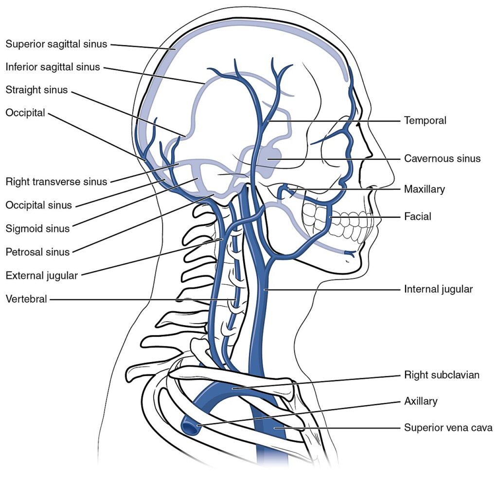
Throughout its course, the plexus communicates superiorly with the basilar and occipital venous plexuses, laterally with the segmental veins, and inferiorly with the pelvic venous plexuses, creating a longitudinal collateral pathway independent of the caval venous system. 

This extensive anatomical continuity establishes the vertebral venous plexus as the principal venous conduit linking the cranial cavity, vertebral column, spinal cord, thorax, abdomen, and pelvis.

ANATOMY

Structural Organization

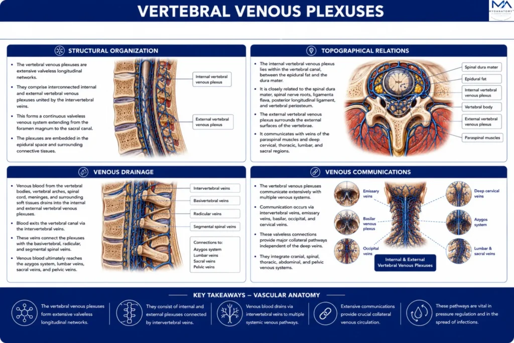
The vertebral venous plexus comprises interconnected internal and external vertebral venous plexuses united by the intervertebral veins, forming a continuous longitudinal valveless venous network extending from the foramen magnumto the sacral canal

The internal vertebral venous plexus occupies the epidural space between the spinal dura mater and the vertebral periosteum, whereas the external vertebral venous plexus surrounds the vertebral bodies, vertebral arches, spinous processes, and adjacent paraspinal musculature. 

Numerous transverse and longitudinal anastomoses create a highly interconnected collateral system that permits multidirectional venous flow throughout the vertebral column.

Exam Question

Describe the structural organization of the vertebral venous plexus, emphasizing the arrangement and anatomical relationships of its internal and external components and the role of the intervertebral veins.

Topographical Relations

The internal vertebral venous plexus lies within the vertebral canal, embedded in the epidural fat and closely related to the spinal dura mater, spinal nerve roots, ligamenta flava, posterior longitudinal ligament, and vertebral periosteum. 

The external vertebral venous plexus surrounds the external surfaces of the vertebrae, communicating with veins of the paraspinal muscles and deep cervical, thoracic, lumbar, and sacral regions. 

Superiorly, the plexus is continuous with the basilar venous plexus and occipital venous sinus, whereas inferiorly it extends into the sacral venous network, establishing uninterrupted craniospinal continuity.

Exam Question

Discuss the topographical relationships of the vertebral venous plexus within and around the vertebral column, highlighting its relationships to the spinal meninges, vertebrae, paraspinal tissues, and craniospinal junction.

Venous Drainage

The vertebral venous plexus receives venous blood from the vertebral bodies, vertebral arches, spinal cord, spinal meninges, vertebral ligaments, paraspinal muscles, and surrounding connective tissues through the basivertebral, radicular, and segmental spinal veins

Venous blood exits the vertebral canal via the intervertebral veins, which communicate with the vertebral, deep cervical, posterior intercostal, lumbar, azygos, hemiazygos, ascending lumbar, lateral sacral, and pelvic veins, establishing multiple collateral pathways independent of the caval venous system

Exam Question

Trace the venous drainage of the vertebral venous plexus, identifying its principal tributaries and explaining how venous return is distributed through the segmental and systemic venous systems.

Venous Communications

The vertebral venous plexus forms one of the most extensive venous communication networks of the body. 

Superiorly, it communicates with the basilar venous plexus, occipital venous sinus, marginal sinus, and dural venous sinuses; laterally, with the vertebral, deep cervical, posterior intercostal, lumbar, and sacral veins; and inferiorly, with the pelvic venous plexuses

Through the intervertebral veins, these communications establish continuous valveless venous continuity between the cranial cavity, vertebral column, thorax, abdomen, and pelvis, creating the principal longitudinal collateral venous pathway of the axial skeleton.

 

Exam Question

Analyze the venous communications of the vertebral venous plexus and explain how its longitudinal valveless architecture integrates the cranial, spinal, thoracic, abdominal, and pelvic venous systems.

SUMMARY TABLE

Scroll to Top