Deep Veins

MYO CORE

Clinical Relevance

The deep craniofacial venous system possesses extensive valveless anastomoses linking the superficial face, orbit, skull base, vertebral column, and intracranial venous sinuses. This unique anatomical organization permits bidirectional venous flow, providing collateral drainage while creating potential pathways for the spread of infection, thrombosis, hemorrhage, and metastatic disease throughout the craniospinal venous system.

OVERVIEW

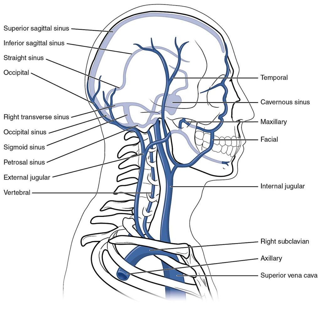
The clinical significance of the deep craniofacial venous system derives from its highly interconnected valveless architecture and extensive communications between the deep facial vein, pterygoid venous plexus, ophthalmic veins, vertebral venous plexus, and dural venous sinuses

Unlike peripheral veins, the absence of valves allows physiological reversal of venous flow according to regional pressure gradients, facilitating collateral venous return but also permitting pathological propagation of infection, thromboembolic material, hemorrhage, and malignant cells. 

These anatomical relationships underlie numerous disorders involving the orbit, skull base, cranial cavity, vertebral column, and deep facial spaces, making detailed knowledge of the deep venous anatomy essential for clinical diagnosis, surgical planning, and radiological interpretation.

 

ANATOMY

Infectious Spread

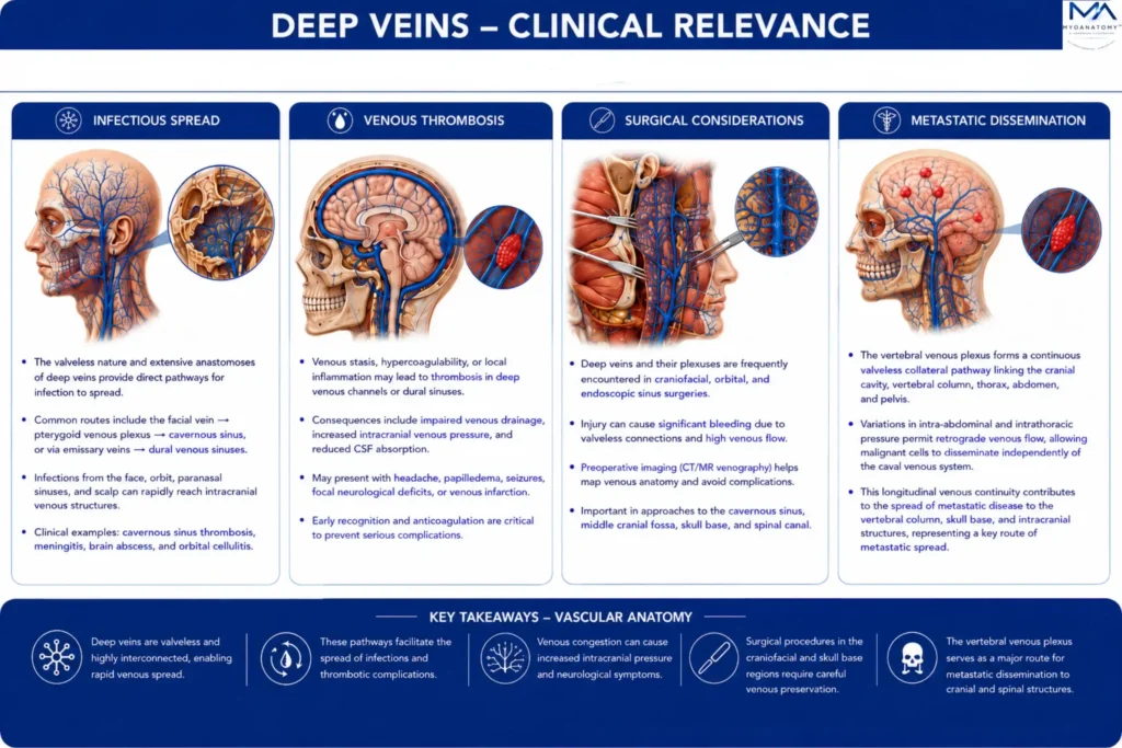
The extensive valveless anastomoses of the deep craniofacial venous system permit bidirectional venous flow, allowing infections originating within the superficial face, deep facial spaces, orbit, paranasal sinuses, nasal cavity, oral cavity, and infratemporal region to extend along normal venous pathways. 

Through the deep facial vein, pterygoid venous plexus, ophthalmic veins, and emissary veins, pathogens may reach the cavernous sinus, adjacent dural venous sinuses, and intracranial compartment without traversing lymphatic barriers. 

Consequently, localized facial infections may progress to life-threatening intracranial complications despite their superficial origin.

Exam Question

Explain how the valveless organization and anatomical communications of the deep craniofacial venous system facilitate the intracranial spread of infection.

Venous Thrombosis

The intimate continuity between the ophthalmic veins, pterygoid venous plexus, emissary veins, and cavernous sinus provides potential pathways for thrombus propagation throughout the craniofacial venous network. 

Venous thrombosis may obstruct normal orbital or intracranial venous drainage, producing venous congestion, orbital edema, increased intracranial venous pressure, and neurological deficits. 

The absence of venous valves permits thrombotic extension in either direction according to regional pressure gradients, contributing to the rapid progression of disease.

Exam Question

Discuss the anatomical basis for venous thrombosis within the deep craniofacial venous system and explain how its valveless architecture influences thrombus propagation.

Metastatic Dissemination

The vertebral venous plexus forms a continuous valveless collateral pathway linking the cranial cavity, vertebral column, thorax, abdomen, and pelvis. 

Variations in intra-abdominal and intrathoracic pressure permit retrograde venous flow, allowing malignant cells to disseminate independently of the caval venous system. 

This longitudinal venous continuity contributes to the spread of metastatic disease to the vertebral column, skull base, and intracranial structures and represents one of the principal anatomical routes of hematogenous skeletal metastasis.

Exam Question

Analyze the role of the vertebral venous plexus in the hematogenous dissemination of metastatic disease and explain the anatomical mechanisms responsible for retrograde venous spread.

Surgical Consideration

The deep craniofacial venous system is closely related to the orbit, infratemporal fossa, skull base, paranasal sinuses, and vertebral canal, making it particularly vulnerable during maxillofacial, orbital, neurosurgical, endoscopic sinus, and skull base procedures. 

The extensive valveless venous communications and marked anatomical variability increase the risk of significant hemorrhage, venous air embolism, and inadvertent intracranial venous injury. 

Detailed knowledge of the deep venous anatomy is therefore essential for surgical planning, radiological interpretation, and the prevention of neurovascular complications.

Exam Question

Evaluate the surgical significance of the deep craniofacial venous system, emphasizing how its anatomy influences operative planning and the prevention of neurovascular complications.

SUMMARY TABLE

Scroll to Top