Deep Veins

MYO CORE

Core Anatomy

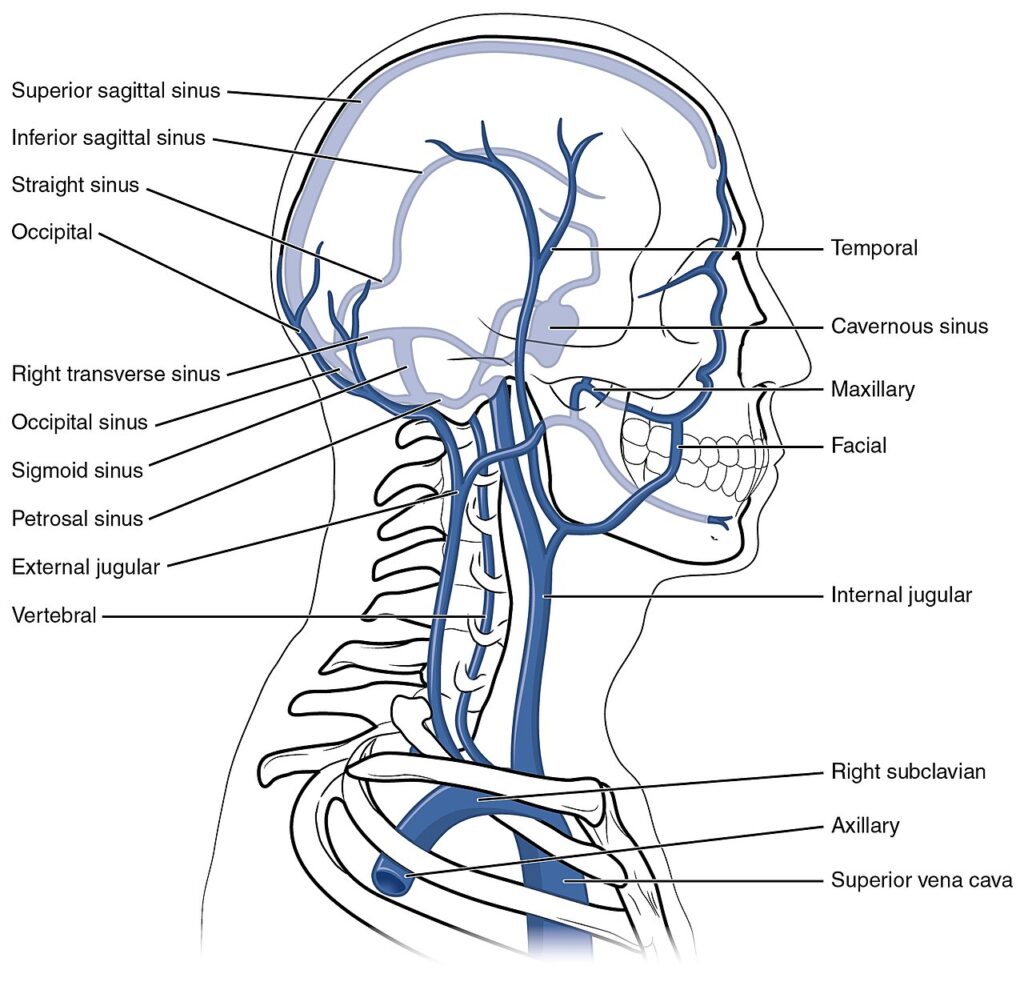
The deep venous system of the head and neck comprises a network of deep veins, venous plexuses, and dural venous sinuses that drain the deep craniofacial structures and upper cervical region. Centered on the deep facial vein, pterygoid venous plexus, ophthalmic veins, pharyngeal venous plexus, vertebral venous plexuses, and internal jugular vein, it forms the principal pathway for deep craniofacial venous return.

OVERVIEW

Rather than following a single linear drainage pattern, venous return is distributed through the deep facial vein, pterygoid venous plexus, superior and inferior ophthalmic veins, pharyngeal venous plexus, vertebral venous plexuses, and dural venous sinuses, creating multiple pathways for regional venous convergence and redistribution. 

These interconnected channels receive blood from the deep facial musculature, orbit, nasal cavity, paranasal sinuses, infratemporal and pterygopalatine fossae, pharynx, meninges, brain, and upper cervical structures before ultimately draining into the internal jugular and vertebral venous systems

The resulting three-dimensional venous architecture provides efficient hemodynamic adaptation, preserves venous outflow during regional obstruction, and integrates the principal drainage territories of the craniovertebral junction, skull base, and deep cervicofacial compartments.

 

ANATOMY

Structural Drainage

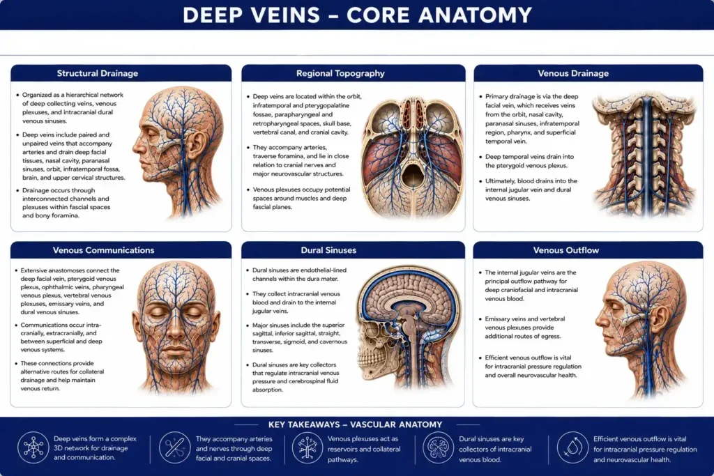
The deep venous system is organized as a hierarchical network of deep collecting veins, venous plexuses, and intracranial dural venous sinuses occupying the deep anatomical compartments of the head and neck. 

Unlike the predominantly linear configuration of the superficial veins, the deep system is characterized by interconnected plexiform architecture, in which multiple venous channels converge, diverge, and intercommunicate within confined fascial and osseous spaces. 

This structural organization provides an integrated framework for venous return from the orbit, deep face, skull base, pharynx, cranial cavity, and upper cervical region while maintaining continuous communication between adjacent deep venous territories.

Exam Question

Explain how the hierarchical organization of the deep venous system differs from the superficial venous system, and discuss the anatomical significance of venous plexuses in coordinating craniofacial venous drainage.

Regional Topography

The deep veins are situated within the orbit, infratemporal and pterygopalatine fossae, parapharyngeal and retropharyngeal spaces, skull base, vertebral canal, and cranial cavity, where they accompany the principal neurovascular structures. 

Their anatomical relationships with the muscles ofmastication, pharyngeal musculature, cranial nerves, major arteries, cervical fasciae, and skull base foramina establish a three-dimensional venous network extending from the facial skeleton to the craniovertebral junction. 

This topographical arrangement integrates the deep fascial compartments into a continuous venous system while preserving regional anatomical continuity.

Exam Question

Describe the three-dimensional topographical relationships of the deep venous system within the orbit, skull base, infratemporal region, pharynx, and cervical compartments, and explain how these relationships integrate the principal neurovascular pathways of the head and neck.

Venous Drainage

The deep venous system receives blood from the orbit, deep facial tissues, nasal cavity, paranasal sinuses, infratemporal region, pharynx, meninges, brain, vertebral canal, and upper cervical structures through a series of interconnected venous channels and plexuses. 

Regional tributaries progressively converge into larger collecting veins, with venous return directed predominantly toward the internal jugular vein and the internal and external vertebral venous plexuses

This hierarchical drainage pattern establishes the principal outflow pathways of the deep craniofacial venous circulation while ensuring efficient integration of intracranial and extracranial drainage territories.

Exam Question

Trace the hierarchical drainage of venous blood from the deep craniofacial structures to the terminal venous outflow pathways, identifying the major collecting veins, venous plexuses, and final drainage into the internal jugular and vertebral venous systems.

Venous Communications

Extensive anastomoses interconnect the deep facial vein, pterygoid venous plexus, superior and inferior ophthalmic veins, pharyngeal venous plexus, vertebral venous plexuses, emissary veins, and dural venous sinuses into a unified three-dimensional venous network

These communications establish anatomical continuity between the orbit, deep face, skull base, cranial cavity, and cervical region, while providing multiple routes for physiological redistribution of venous blood. 

The resulting anastomotic architecture integrates the principal deep venous territories into a coordinated craniovertebral drainage system and represents the defining structural characteristic of the deep craniofacial venous network.

Exam Question

Analyze the anatomical communications between the deep facial vein, pterygoid venous plexus, ophthalmic veins, emissary veins, vertebral venous plexuses, and dural venous sinuses, and explain how these interconnections establish a unified craniovertebral venous network.

SUMMARY TABLE

Scroll to Top