Deep Veins
MYO CORE
Ophthalmic Veins
The ophthalmic veins comprise the superior and inferior ophthalmic veins, forming the principal valveless venous system of the orbit. They provide the primary orbital venous outflow to the cavernous sinus while communicating with the facial vein, pterygoid venous plexus, and intracranial venous channels, thereby integrating the orbital, facial, deep craniofacial, and intracranial venous systems.
OVERVIEW
The superior and inferior ophthalmic veins constitute the principal valveless venous drainage system of the orbit, coursing through the intraconal and extraconal compartments in close association with the orbital neurovascular structures.
The superior ophthalmic vein, the dominant orbital vein, originates near the medial angle of the orbit from the union of the supraorbital and angular veins, courses posteriorly through the superior orbit, and terminates in the cavernous sinus via the superior orbital fissure.
The inferior ophthalmic vein drains the inferior orbit and commonly terminates in the cavernous sinus, the superior ophthalmic vein, or the pterygoid venous plexus. Together, the ophthalmic veins receive tributaries from the central retinal vein, vorticose veins, anterior ciliary veins, lacrimal veins, muscular veins, ethmoidal veins, and palpebral and conjunctival veins, integrating venous drainage from the globe, orbit, and adjacent orbital structures.
Their extensive valveless anastomoses establish continuous venous communication between the orbit, superficial face, deep craniofacial compartments, skull base, and intracranial venous system.

ANATOMY
Formation & Origin
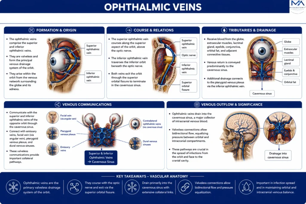
The superior ophthalmic vein is the principal orbital vein, typically originating at the superomedial angle of the orbit from the union of the supraorbital vein and the angular vein, although its formation may vary owing to the highly anastomotic orbital venous system.
The inferior ophthalmic vein arises as a plexiform network within the floor and lateral aspect of the orbit through the convergence of veins draining the inferior orbital structures.
Together, these vessels establish the principal valveless venous outflow of the orbit and constitute the anatomical foundation of the orbital venous system.
Exam Question
Describe the formation of the superior and inferior ophthalmic veins, identifying their typical origins and explaining their contribution to the organization of the orbital venous system.
Course & Relations
The superior ophthalmic vein courses posteriorly through the superior orbit, passing superior to the optic nerve while accompanying the ophthalmic artery within the orbital fat. It exits the orbit through the superior orbital fissure to terminate in the cavernous sinus.
The inferior ophthalmic vein traverses the inferior orbital compartment beneath the optic nerve and demonstrates variable termination, draining either directly into the cavernous sinus, joining the superior ophthalmic vein, or communicating with the pterygoid venous plexus through the inferior orbital fissure. Throughout their course, both veins remain closely related to the extraocular muscles, orbital fascia, and orbital neurovascular structures.
Exam Question
Trace the course of the superior and inferior ophthalmic veins from their origins to their terminations, emphasizing their anatomical relationships within the orbit and at the orbital apex.
Tributaries & Drainage
The ophthalmic veins receive venous blood from virtually all orbital structures through numerous tributaries. Major tributaries include the central retinal vein, vorticose (vortex) veins, anterior ciliary veins, lacrimal veins, muscular veins, ethmoidal veins, palpebral veins, and conjunctival veins.
Collectively, these vessels drain the globe, extraocular muscles, lacrimal gland, eyelids, conjunctiva, orbital fat, and adjacent connective tissues.
Venous return is conveyed predominantly to the cavernous sinus, with additional drainage to the pterygoid venous plexus through the inferior ophthalmic vein, integrating the orbital circulation with the deep craniofacial venous system.
Exam Question
Identify the principal tributaries of the ophthalmic veins and explain the hierarchical pathway of venous drainage from the orbital contents to the cavernous sinus and pterygoid venous plexus.
Venous Communications
The ophthalmic veins form one of the principal venous communication systems of the head through extensive valveless anastomoses.
Anteriorly, they communicate with the facial vein through the angular vein; inferiorly, the inferior ophthalmic vein communicates with the pterygoid venous plexus via the inferior orbital fissure; posteriorly, both veins communicate with the cavernous sinus, which in turn connects with the remaining dural venous sinuses.
Additional communications with the central retinal, ethmoidal, and orbital venous networks create a continuous pathway linking the orbit with the superficial face, deep craniofacial compartments, skull base, and intracranial venous circulation.
Exam Question
Analyze the venous communications of the ophthalmic veins and discuss how these anatomical connections integrate the orbital, facial, deep craniofacial, skull base, and intracranial venous systems.
SUMMARY TABLE