Dural Sinuses

MYO CORE

Core Anatomy

The dural venous sinuses are rigid, endothelial-lined, valveless venous channels formed between the periosteal and meningeal layers of the cranial dura mater. Unlike conventional veins, they possess no smooth muscle or valves and remain permanently patent because of their dense fibrous dural walls.

OVERVIEW

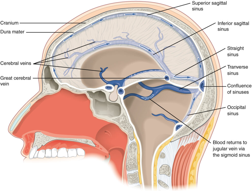
Dural Venous Sinuses collect venous blood from the brain, cerebellum, meninges, orbit, skull, and diploë while serving as the principal site of cerebrospinal fluid (CSF) absorption through arachnoid granulations. 

Together, the dural venous sinuses form an integrated, valveless intracranial venous network embedded within the dura mater

Their rigid architecture permits pressure-dependent multidirectional blood flow, ensuring efficient cerebral venous drainage while providing extensive collateral communication with the cavernous sinus, ophthalmic veins, emissary veins, vertebral venous plexuses, and internal jugular veins. 

By integrating cerebral venous outflow with continuous CSF resorption, they maintain intracranial pressure, cerebral hemodynamic homeostasis, and craniospinal venous continuity.

ANATOMY

Dural Formation

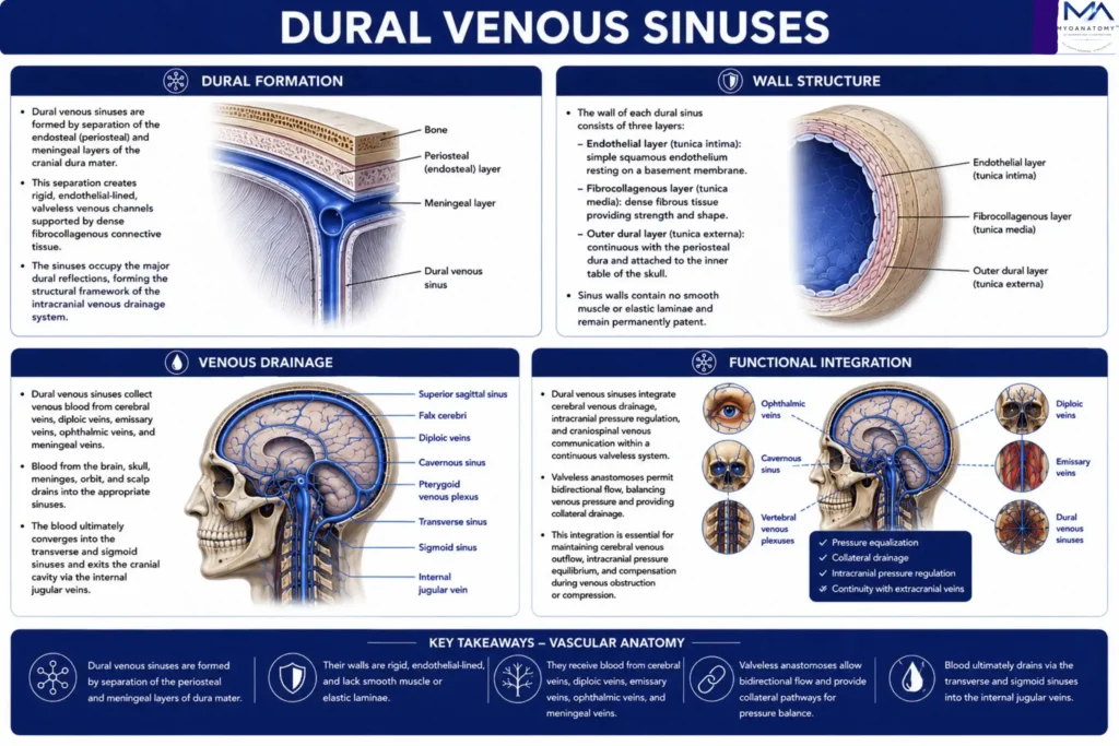
The dural venous sinuses are formed by separation of the endosteal (periosteal) and meningeal layers of the cranial dura mater, creating rigid endothelial-lined venous channels supported by dense fibrocollagenous connective tissue. 

Unlike conventional veins, their walls contain neither smooth muscle nor elastic lamina and therefore remain permanently patent, resisting luminal collapse despite physiological fluctuations in intracranial pressure. 

The sinuses occupy the major dural reflections-including the falx cerebri, tentorium cerebelli, falx cerebelli, and diaphragma sellaeforming the structural framework of the intracranial venous drainage system.


Exam Question

Explain how the anatomical relationship between the endosteal and meningeal dural layers determines the structural stability, topographical distribution, and permanent patency of the dural venous sinuses.

Wall Structure

Each dural venous sinus is lined by a continuous simple squamous endothelium resting directly upon dense fibrocollagenous dura without an intervening tunica media, elastic lamina, or venous valves. 

The outer boundary is formed by the endosteal dura firmly adherent to the inner calvaria,whereas the inner boundary consists of the meningeal dura. 

Regions containing arachnoid granulations penetrate the endothelial lining to facilitate pressure-dependent, unidirectional transfer of cerebrospinal fluid into the venous circulation while preserving intracranial pressure homeostasis.


Exam Question

Describe the histological organization of the dural venous sinus wall and explain how its specialized endothelial architecture permits simultaneous cerebral venous drainage and cerebrospinal fluid resorption.

Venous Drainage

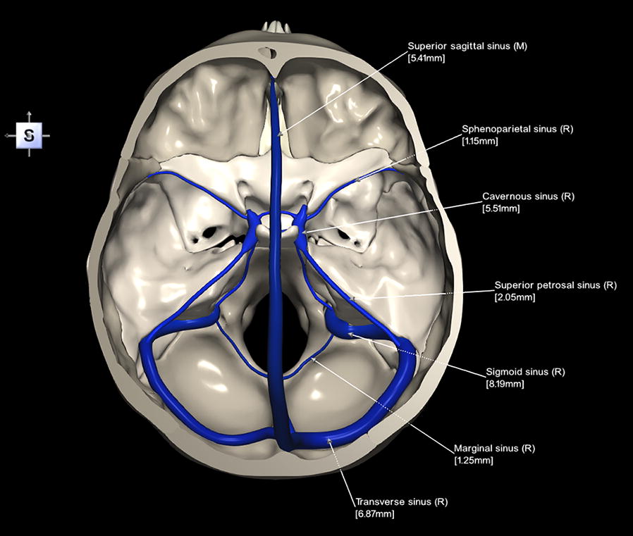
The dural venous sinuses receive venous return from the superficial and deep cerebral veins, cerebellar veins, diploic veins, meningeal veins, ophthalmic veins, emissary veins, and the cavernous sinus.

Because the system is entirely valveless, venous blood redistributes according to dynamic pressure gradients generated by cerebral perfusion, respiration, and posture, allowing extensive collateral flow. 

Venous drainage ultimately converges through the transverse and sigmoid sinuses, which exit the cranial cavity via the jugular foramina as the internal jugular veins, constituting the principal pathway of cerebral venous outflow.

Exam Question

Trace the hierarchical drainage of venous blood from the deep craniofacial structures to the terminal venous outflow pathways, identifying the major collecting veins, venous plexuses, and final drainage into the internal jugular and vertebral venous systems.

Functional Integration

The dural venous sinuses integrate cerebral venous drainage, cerebrospinal fluid resorption, intracranial pressure regulation, and craniospinal venous communication within a continuous endothelial network. 

Extensive anastomoses with the ophthalmic veins, emissary veins, pterygoid venous plexus, vertebral venous plexuses, and extracranial veins preserve cerebral venous return during obstruction while simultaneously providing potential routes for intracranial propagation of infection, thrombosis, neoplastic dissemination, and hemorrhagic extension. 

Consequently, the dural sinus system represents a fundamental interface between cerebral physiology, skull-base anatomy, neuroradiology, and neurosurgical practice.


Exam Question

Critically analyze how the structural organization and valveless collateral architecture of the dural venous sinuses optimize cerebral venous homeostasis while facilitating intracranial spread of infection, thrombosis, and metastatic disease.

SUMMARY TABLE

Scroll to Top