Dural Sinuses

MYO CORE

Clinical Relevance

The clinical significance of the intracranial venous system is determined by its rigid endothelial dural channels, absence of venous valves, extensive collateral communications, and intimate relationships with the brain, cranial nerves, orbit, skull base, and cerebrospinal fluid pathways

OVEVIEW

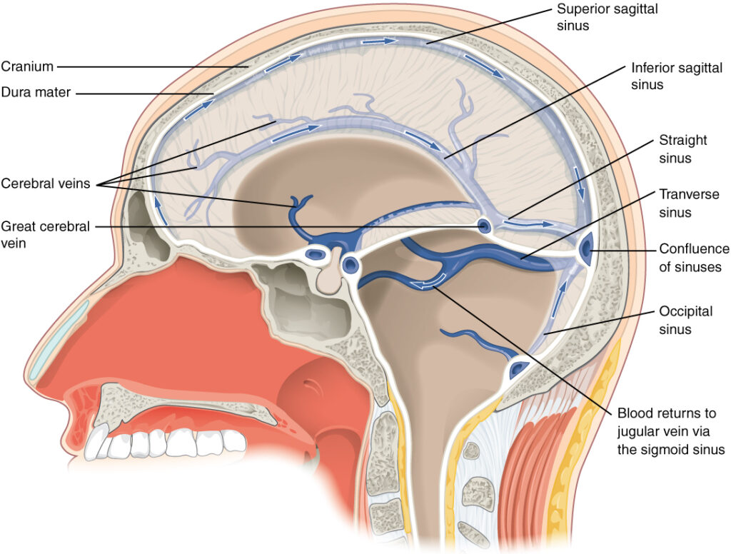
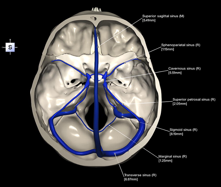
The dural venous sinuses and their extracranial venous communications form a highly integrated valveless collateral drainage network that extends from the cranial cavity to the orbit, face, skull base, neck, and vertebral venous system

Beyond providing cerebral venous outflow, this network maintains cerebral venous return, cerebrospinal fluid (CSF) resorption through the arachnoid granulations, intracranial pressure homeostasis, and compensatory redistribution of venous blood during physiological pressure fluctuations or venous obstruction. 

Owing to its extensive collateral communications and absence of venous valves, pathological processes-including venous thrombosis, intracranial hypertension, traumatic injury, skull-base fractures, infection, and metastatic disseminationmay rapidly propagate throughout the craniospinal venous system. 

Consequently, precise anatomical understanding combined with CT and MR venography is essential for accurate diagnosis, neurosurgical planning, endovascular intervention, and postoperative evaluation.

ANATOMY

Venous Thrombosis

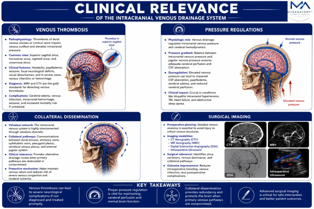
The valveless architecture of the dural venous sinuses permits unrestricted pressure-dependent venous flow but also facilitates propagation of thrombus throughout the intracranial venous network. 

Cavernous sinus thrombosis typically results from septic spread through the facial, angular, ophthalmic, or pterygoid venous pathways, whereas dural venous sinus thrombosis (DVST) most frequently involves the superior sagittal, transverse, or sigmoid sinuses

Progressive venous obstruction elevates cerebral venous pressure, impairs cerebrospinal fluid absorption, reduces cerebral perfusion, and disrupts the blood-brain barrier, producing vasogenic edema, venous infarction, intracerebral hemorrhage, seizures, cranial neuropathies, and potentially fatal cerebral herniation.

Exam Question

Compare the anatomical pathways, mechanisms of propagation, and cerebrovascular consequences of cavernous sinus thrombosis and dural venous sinus thrombosis, emphasizing the role of valveless venous anatomy in disease progression.

Pressure Regulations

The arachnoid granulations project directly into the superior sagittal and adjacent dural venous sinuses, making cerebrospinal fluid absorption dependent upon a favorable pressure gradient between the subarachnoid space and the venous sinuses

Venous thrombosis, congenital sinus stenosis or hypoplasia, external compression, or elevated central venous pressure diminish this gradient, impairing CSF resorption and producing intracranial hypertension

Progressive elevation of intracranial pressure may result in papilledema, headache, visual impairment, reduced cerebral perfusion, transtentorial herniation, and irreversible neurological injury, although collateral venous pathways may temporarily compensate for impaired primary drainage.

Exam Question

Explain how the anatomical relationship between the dural venous sinuses and arachnoid granulations governs cerebrospinal fluid dynamics and analyze the mechanisms by which venous outflow obstruction produces intracranial hypertension.

Collateral Dissimenation

Extensive communications between the ophthalmic veins, emissary veins, pterygoid venous plexus, vertebral venous plexuses, and dural venous sinuses establish a continuous valveless collateral network linking the cranial cavity with the orbit, face, scalp, skull base, neck, and vertebral canal. 

Although these pathways preserve cerebral venous drainage during physiological adaptation or venous obstruction, they also permit retrograde dissemination of infection, septic thrombi, inflammatory mediators, air emboli, and metastatic tumor cells between intracranial and extracranial compartments. 

This anatomical organization underlies the rapid spread offacial infections to the cavernous sinus ( one way via  angular vein) , vertebral metastatic dissemination, and the characteristic patterns of craniospinal venous disease.


Exam Question

Critically evaluate how the collateral anatomy of the intracranial venous system simultaneously preserves cerebral venous homeostasis while facilitating the craniospinal dissemination of infection, thrombosis, air emboli, and metastatic disease.

Surgical Imaging

The dural venous sinuses are critical surgical landmarks because of their close relationships with the calvaria, skull base, cranial nerves, and major intracranial vessels. Sinus injury during trauma or cranial and skull-base surgery may cause massive venous hemorrhage, venous infarction, air embolism, or impaired cerebral venous drainage

CT venography (CTV) is the primary modality for detecting acute sinus injury, skull-base fractures, and thrombosis, whereas MR venography (MRV) better demonstrates venous patency, anatomical variants, collateral circulation, and chronic venous disease. 

Together, detailed anatomical knowledge and advanced venous imaging are essential for diagnosis, surgical planning, neurovascular intervention, and postoperative assessment

Exam Question

Analyze how the anatomical relationships of the dural venous sinuses and the complementary roles of CT and MR venography optimize the diagnosis, surgical planning, and management of intracranial venous disorders.

SUMMARY TABLE

Scroll to Top