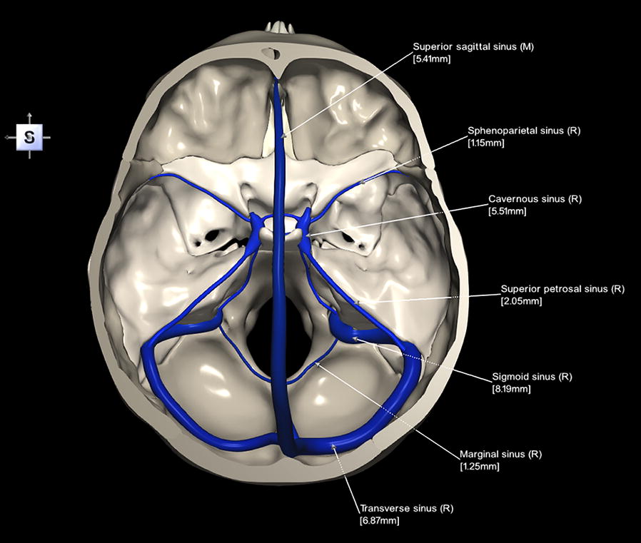
Dural Sinuses
MYO CORE
Venous Drainage
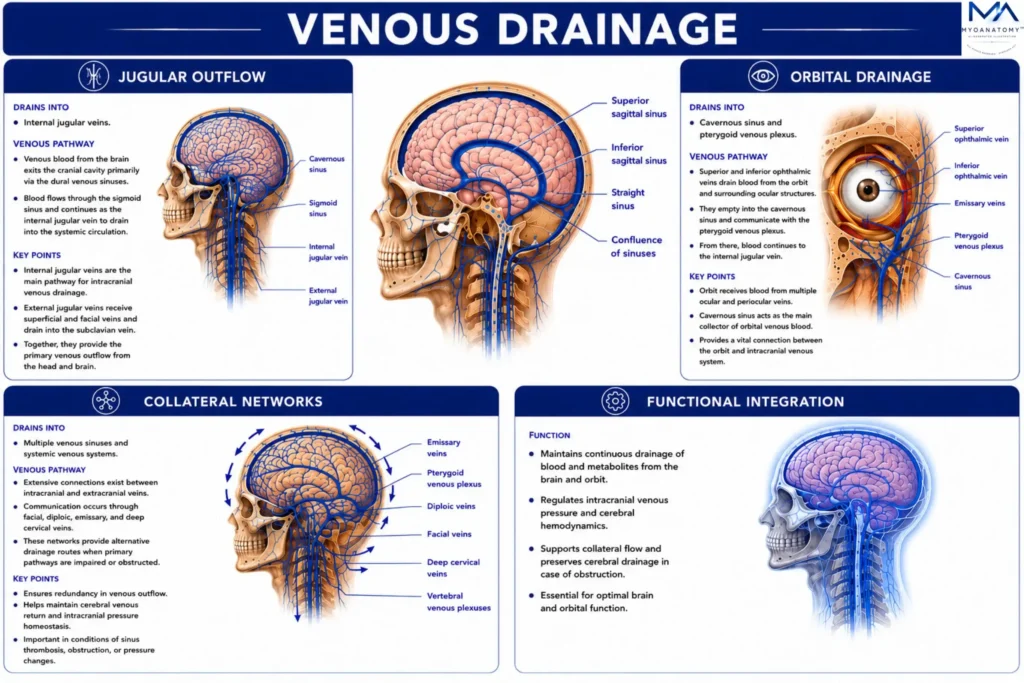
The intracranial venous drainage system comprises the internal jugular veins, ophthalmic veins, emissary veins, pterygoid venous plexus, vertebral venous plexuses, and extensive extracranial venous communications, forming an integrated valveless collateral network.
OVERVIEW
The intracranial venous drainage network extends beyond the dural venous sinuses through an extensive system of valveless extracranial communications that ensures uninterrupted cerebral venous outflow.
Venous blood exits the cranial cavity primarily through the internal jugular veins, while the ophthalmic veins, emissary veins, pterygoid venous plexus, vertebral venous plexuses, and superficial craniofacial veins establish multidirectional collateral pathways linking the cranial cavity with the orbit, face, skull base, neck, and vertebral canal.
Collectively, these interconnected channels redistribute venous return according to regional pressure gradients, preserving cerebral venous homeostasis, intracranial pressure equilibrium, cerebral hemodynamic stability, and functional continuity between the intracranial, craniofacial, cervical, and vertebral venous systems.

ANATOMY
Jugular Outflow
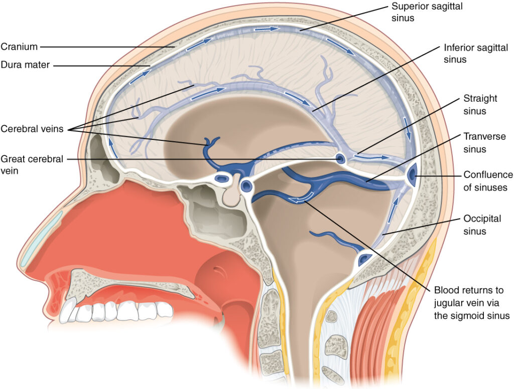
The internal jugular vein originates as the direct continuation of the sigmoid sinus at the jugular foramen, where it expands into the superior jugular bulb and receives the inferior petrosal sinus.
Descending within the carotid sheath, it receives multiple extracranial tributaries before uniting with the subclavian vein to form the brachiocephalic vein.
As the principal venous outflow pathway of the cranial cavity, it conveys the majority of cerebral venous return from the dural venous sinuses to the systemic circulation while maintaining pressure-dependent continuity with collateral venous channels.
Exam Question
Explain how the anatomical formation, topographical relationships, tributaries, and termination of the internal jugular vein establish it as the principal venous outflow pathway of the cranial cavity.
Orbital Drainage
The superior and inferior ophthalmic veins provide direct valveless communication between the orbit and the cavernous sinus, while the angular, facial, and deep facial veins establish continuity with the extracranial venous system.
These vessels receive venous blood from the orbital contents, eyelids, and adjacent facial structures before draining into the cavernous sinus or pterygoid venous plexus.
Their absence of valves permits multidirectional venous flow, preserving collateral drainage while simultaneously providing potential pathways for intracranial extension of infection, thrombosis, and inflammatory disease.
Exam Question
Analyze how the anatomical organization and venous communications of the ophthalmic veins integrate orbital, facial, and intracranial venous drainage while facilitating both collateral circulation and pathological spread.
Collateral Networks
The emissary veins traverse cranial foramina to connect the dural venous sinuses with the extracranial scalp veins, whereas the pterygoid venous plexus forms an extensive venous network within the infratemporal fossa that communicates with the cavernous sinus through emissary and ophthalmic veins.
Posteriorly, the internal and external vertebral venous plexuses establish uninterrupted valveless continuity with the basilar venous plexus, occipital sinus, cervical veins, and pelvic venous system, extending from the cranial cavity to the sacrum.
Together, these interconnected channels constitute the principal collateral pathways between the cranial cavity, skull base, vertebral canal, and systemic venous circulation.
Exam Question
Evaluate how emissary veins, the pterygoid venous plexus, and the vertebral venous plexuses establish an integrated collateral network linking the cranial cavity with the extracranial and spinal venous systems.
Functional integration
The intracranial venous drainage system functions as a dynamic valveless collateral network, integrating the internal jugular veins, ophthalmic veins, emissary veins, pterygoid venous plexus, vertebral venous plexuses, superficial craniofacial veins, and dural venous sinuses into a single hemodynamic unit.
Continuous redistribution of venous blood through these interconnected pathways maintains cerebral venous return during postural change, respiration, and venous obstruction while preserving intracranial pressure homeostasis.
The same collateral architecture, however, provides anatomical routes for the intracranial dissemination of infection, septic thrombosis, air emboli, and metastatic disease, highlighting the close relationship between normal venous physiology and clinical pathology.
Exam Question
Critically analyze how the valveless collateral architecture of the intracranial venous drainage system simultaneously preserves cerebral venous homeostasis and facilitates the craniospinal spread of infection, thrombosis, air emboli, and metastatic disease.
SUMMARY TABLE