Dural Sinuses

MYO CORE

Midline Sinuses

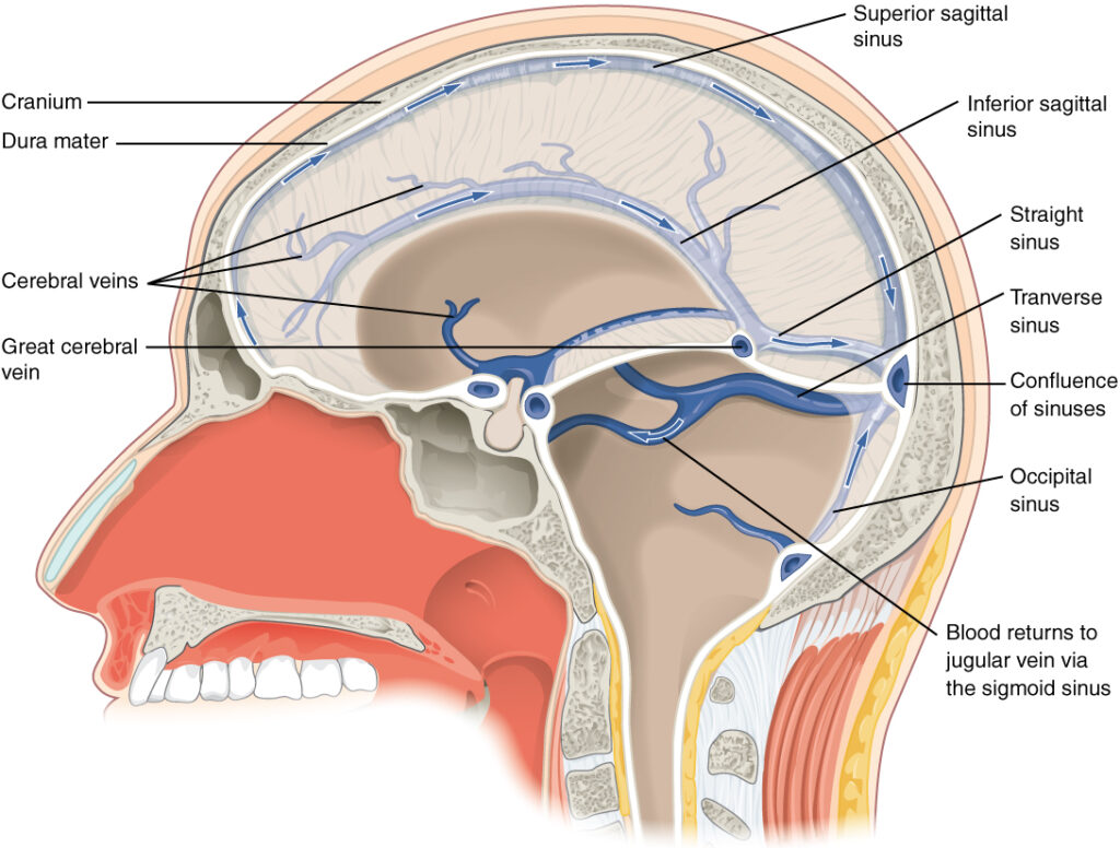
The midline dural venous sinuses comprise the superior sagittal, inferior sagittal, straight, occipital, and confluence of sinuses, forming the principal longitudinal venous drainage axis of the cranial cavity. Embedded within the major dural reflections, they receive venous blood from the superficial and deep cerebral hemispheres, cerebellum, meninges, diploë, and posterior cranial fossa while serving as the primary site of cerebrospinal fluid (CSF) resorption through arachnoid granulations

OVERVIEW

The midline sinuses constitute the central axial framework of the intracranial dural venous system, extending through the falx cerebri, tentorium cerebelli, falx cerebelli, and internal occipital crest to establish uninterrupted venous continuity from the frontal cranial vault to the posterior cranial fossa. 

Together, they integrate superficial cortical, deep cerebral, cerebellar, and posterior fossa venous drainage before directing blood into the transverse sinuses. 

Their rigid, valveless endothelial channels permit multidirectional pressure-dependent venous flow while facilitating continuous cerebrospinal fluid (CSF) resorption through arachnoid granulations and extensive collateral communication with the vertebral venous plexuses. 

Consequently, they preserve cerebral hemodynamic stability, intracranial pressure homeostasis, and functional continuity between the intracranial and spinal venous systems.

ANATOMY

Superior Sagittal Sinus

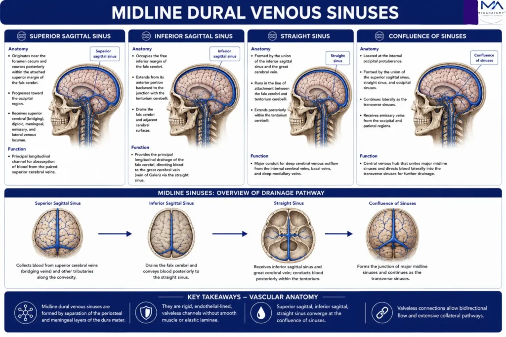
The superior sagittal sinus originates near the foramen cecum and courses posteriorly within the attached superior margin of the falx cerebri, progressively enlarging toward the occipital region. 

Throughout its course, it receives superior cerebral (bridging) veins, diploic veins, meningeal veins, emissary veins, and lateral venous lacunae, into which numerous arachnoid granulations project to facilitate pressure-dependent cerebrospinal fluid resorption.

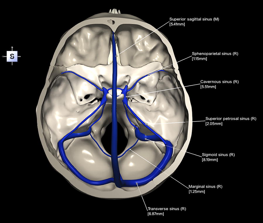
Posteriorly, it usually terminates within the confluence of sinuses, although venous outflow most commonly continues into the dominant right transverse sinus

As the principal collector of the superficial cerebral venous system, it integrates cortical, calvarial, and meningeal drainage while contributing substantially to intracranial pressure regulation.


Exam Question

Explain how the topographical course, tributaries, lateral venous lacunae, and arachnoid granulations of the superior sagittal sinus optimize superficial cerebral venous drainage and cerebrospinal fluid resorption.

Inferior Sagittal Sinus

The inferior sagittal sinus occupies the free inferior margin of the falx cerebri, extending from its anterior portion toward the junction with the tentorium cerebelli. 

Considerably smaller than the superior sagittal sinus, it receives venous blood primarily from the medial cerebral hemispheres, falcine veins, and adjacent deep white matter before uniting posteriorly with the great cerebral vein to form the straight sinus

Functionally, it provides the principal longitudinal drainage pathway of the medial cerebral surfaces and represents the anatomical interface between superficial midline drainage and the deep cerebral venous system.

Exam Question

Describe the anatomical course, tributaries, termination, and functional significance of the inferior sagittal sinus in integrating medial cerebral venous return with the deep cerebral venous circulation.

Straight Sinus

The straight sinus is formed by the union of the inferior sagittal sinus and the great cerebral vein, occupying the line of attachment between the falx cerebri and tentorium cerebelli

It constitutes the principal outflow channel of the deep cerebral venous system, transmitting venous blood from the internal cerebral veins, basal veins, and deep medullary veins conveyed through the great cerebral vein before terminating at the confluence of sinuses

Positioned at the junction of the supratentorial and infratentorial compartments, the straight sinus represents the major anatomical conduit linking deep cerebral drainage with the posterior dural venous system.

Exam Question

Analyze how the anatomical formation, topographical relationships, and venous connections of the straight sinus establish it as the principal outflow pathway of the deep cerebral venous system.

Confluence of Sinuses

The confluence of sinuses (torcular Herophili) is situated adjacent to the internal occipital protuberance, where the superior sagittal, straight, and occipital sinuses converge before distributing venous blood into the paired transverse sinuses

The occipital sinus, located within the attached margin of the falx cerebelli along the internal occipital crest, extends inferiorly toward the foramen magnum, where it communicates with the marginal sinus and the internal vertebral venous plexus

Although frequently the smallest dural venous sinus, it provides an important collateral pathway linking intracranial and spinal venous systems, particularly when the principal cranial venous channels are compromised.

Exam Question

Evaluate how the anatomical organization, physiological asymmetry, and collateral communications of the confluence of sinuses and occipital sinus preserve cerebral venous outflow during both normal physiology and pathological venous obstruction.

SUMMARY TABLE

Scroll to Top