Dural Sinuses
MYO CORE
Lateral & Cavernous Sinuses
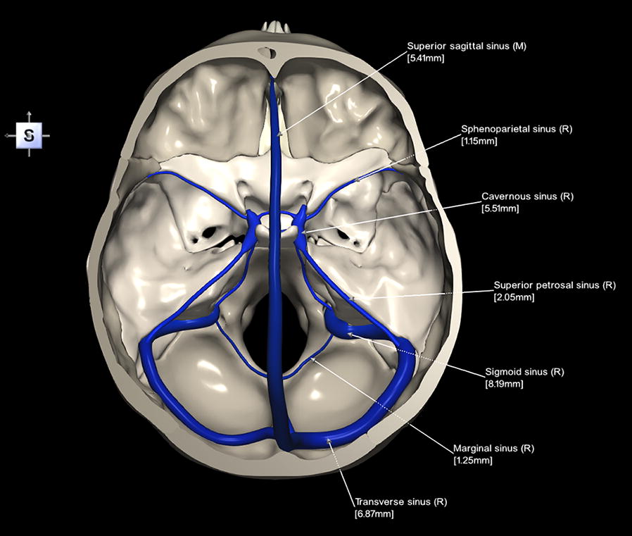
The lateral and cavernous dural venous sinuses comprise the cavernous, transverse, sigmoid, superior petrosal, inferior petrosal, sphenoparietal, intercavernous sinuses, and basilar venous plexus, forming the principal lateral and skull base venous drainage network.
OVERVIEW
The lateral and cavernous sinuses constitute the principal venous channels of the middle and posterior cranial fossae.
Extending along the tentorium cerebelli, petrous temporal bone, sphenoid bone, clivus, and sella turcica, they establish an interconnected valveless network linking the cerebral hemispheres, cerebellum, brainstem, orbit, pituitary gland, face, and vertebral venous system.
Collectively, they receive venous blood from these regions before directing it to the internal jugular veins and vertebral venous plexuses through the cavernous, petrosal, intercavernous, and basilar communications.
Their rigid endothelial architecture permits multidirectional pressure-dependent venous flow, preserving cerebral venous redistribution, orbital and skull base drainage, intracranial pressure homeostasis, and craniospinal venous continuity.

ANATOMY
Cavernous Complex
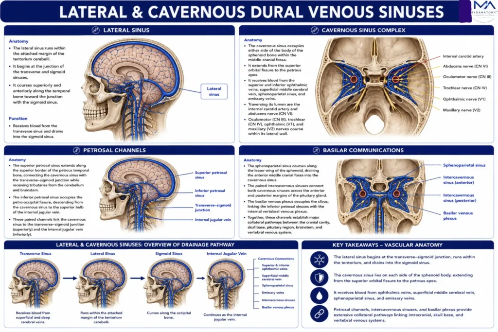
The cavernous sinus occupies either side of the body of the sphenoid bone within the middle cranial fossa, extending from the superior orbital fissure to the petrous apex.
It receives blood from the superior and inferior ophthalmic veins, superficial middle cerebral vein, sphenoparietal sinus, and emissary veins, while communicating extensively with the facial vein, pterygoid venous plexus, petrosal sinuses, and intercavernous sinuses.
Traversing its lumen are the internal carotid artery and abducens nerve (CN VI), whereas the oculomotor (CN III), trochlear (CN IV), ophthalmic (V1), and maxillary (V2) nerves course within its lateral wall.
This unique neurovascular organization establishes the cavernous sinus as the principal venous crossroads between the orbit, face, skull base, and intracranial cavity.
Exam Question
Analyze how the topographical relationships, tributaries, neurovascular contents, and extensive communications of the cavernous sinus explain its central role in intracranial venous drainage and skull base pathology.
Lateral Drainage
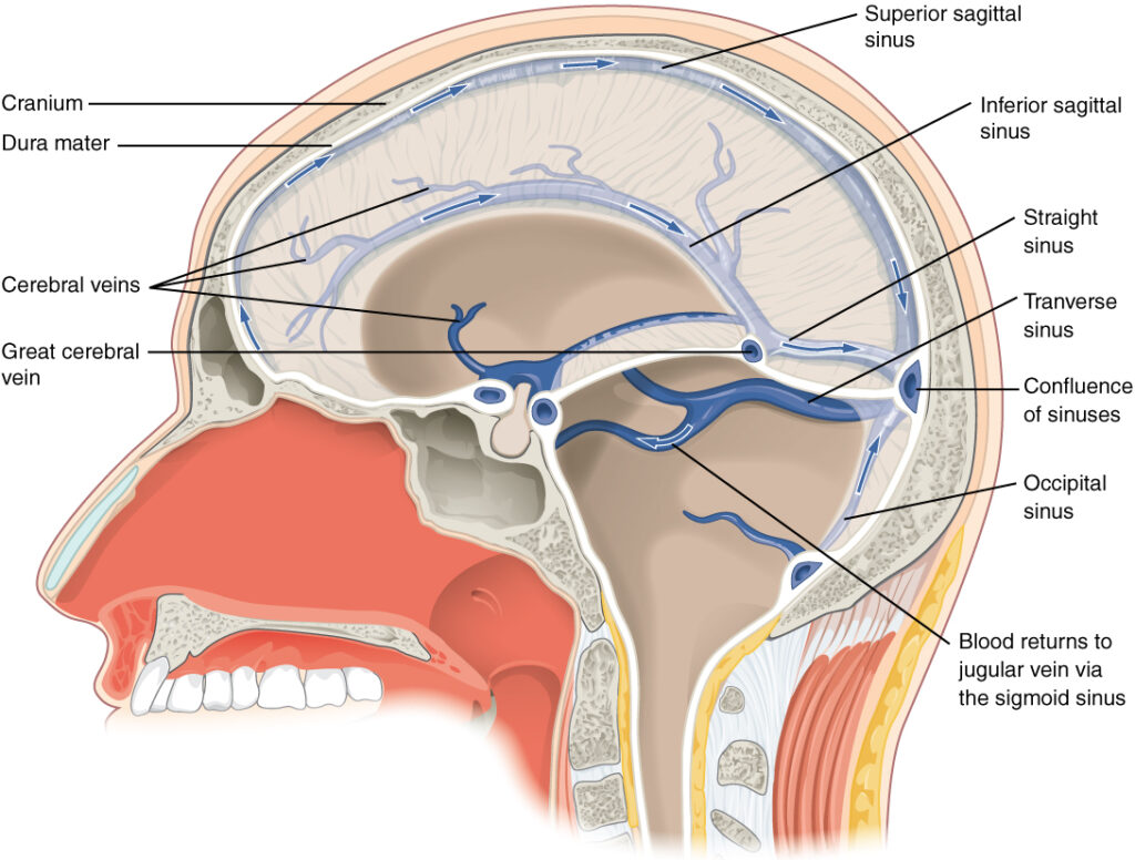
The transverse sinuses arise from the confluence of sinuses, coursing laterally within the attached margin of the tentorium cerebelli along the occipital bone before becoming the sigmoid sinuses.
They receive venous return from the superior sagittal, straight, occipital, superior petrosal, cerebellar, diploic, and meningeal veins.
The sigmoid sinuses descend through the posterior cranial fossa in an S-shaped groove, receiving additional mastoid emissary and cerebellar tributaries before exiting the skull through the jugular foramen, where they continue as the internal jugular veins.
Together, they constitute the principal terminal outflow pathway of the intracranial dural venous system.
Exam Question
Describe the anatomical course, tributaries, and termination of the transverse and sigmoid sinuses, explaining why they represent the principal terminal pathway of intracranial venous drainage.
Petrosal Channels
The superior petrosal sinus extends along the superior border of the petrous temporal bone, connecting the cavernous sinus with the transverse-sigmoid junction while receiving tributaries from the cerebellum and brainstem.
The inferior petrosal sinus occupies the petro-occipital fissure, descending from the cavernous sinus directly to the superior bulb of the internal jugular vein, forming the shortest venous route between the cavernous sinus and systemic circulation.
Together, these paired channels provide the principal venous egress from the cavernous sinus while establishing critical communications between the middle and posterior cranial fossae.
Exam Question
Compare the anatomical relationships, venous connections, and functional significance of the superior and inferior petrosal sinuses in draining the cavernous sinus.
Basilar Communications
The sphenoparietal sinus courses along the lesser wing of the sphenoid, draining the anterior middle cranial fossa into the cavernous sinus.
The paired intercavernous sinuses connect both cavernous sinuses across the anterior and posterior margins of the pituitary gland, forming the circular venous sinus.
Posteriorly, the basilar venous plexus occupies the clivus, linking the inferior petrosal sinuses with the internal vertebral venous plexus.
Together, these channels establish major collateral pathways between the cranial cavity, skull base, pituitary region, brainstem, and vertebral venous system, preserving venous drainage during physiological variation or pathological obstruction.
Exam Question
Analyze how the anatomical organization and communications of the sphenoparietal sinus, intercavernous sinuses, and basilar venous plexus establish collateral venous continuity between the cranial cavity and vertebral venous system.
SUMMARY TABLE