Deep Veins

MYO CORE

Pterygoid Venous Plexus

The pterygoid venous plexus is an extensive three-dimensional valveless plexiform network occupying the infratemporal fossa around the pterygoid muscles and the second part of the maxillary artery. It forms the principal deep venous convergence of the craniofacial region, draining the deep face into the maxillary vein while linking the superficial, deep, orbital, and intracranial venous systems.

OVERVIEW

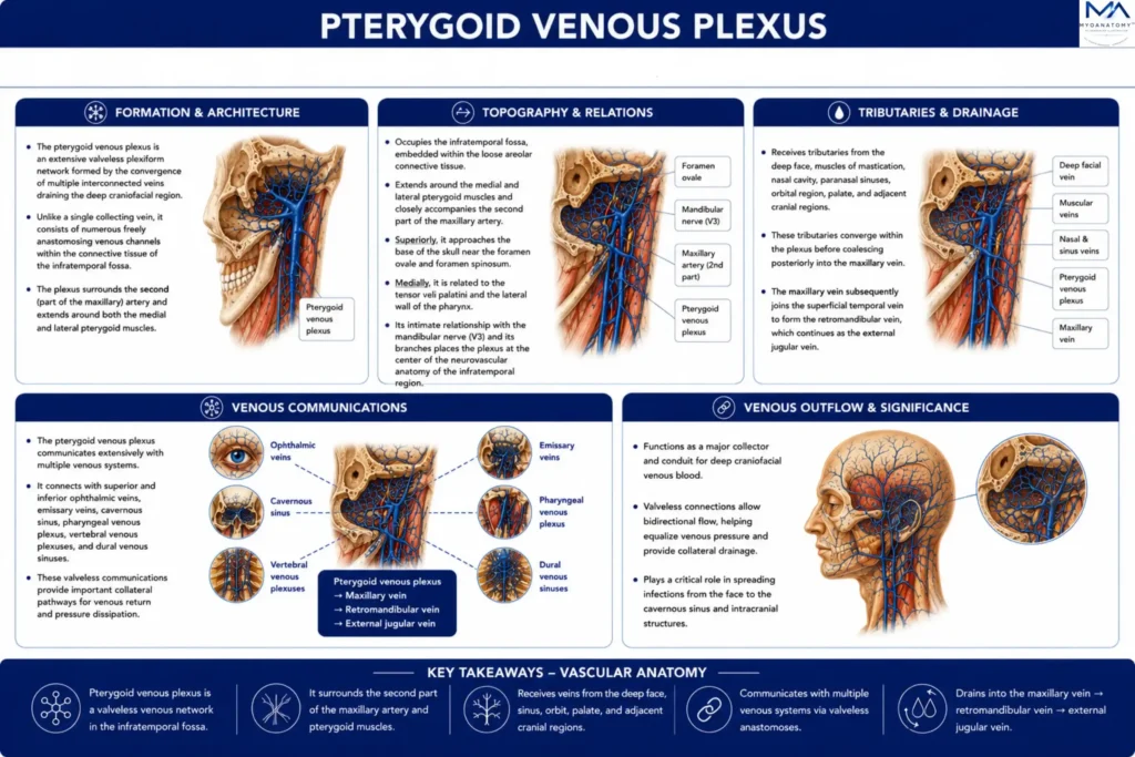
The pterygoid venous plexus consists of numerous freely anastomosing valveless veins embedded within the connective tissue of the infratemporal fossa, surrounding the medial and lateral pterygoid muscles and closely accompanying the second part of the maxillary artery

Its plexiform arrangement occupies the spaces between the muscles of mastication and adjacent neurovascular structures, creating a highly interconnected venous network rather than a single collecting vessel. 

The plexus receives tributaries from the deep face, muscles of mastication, nasal cavity, paranasal sinuses, palate, and adjacent craniofacial regions before converging posteriorly to form the maxillary vein

This hierarchical organization establishes the pterygoid venous plexus as the central venous convergence of the deep craniofacial circulation and the anatomical foundation for its extensive communications with both extracranial and intracranial venous pathways.

ANATOMY

Formation & Architecture

The pterygoid venous plexus is an extensive valveless plexiform network formed by the convergence of multiple interconnected veins draining the deep craniofacial region. 

Unlike a single collecting vein, it consists of numerous freely anastomosing venous channels that create a three-dimensional vascular meshwork embedded within the connective tissue of the infratemporal fossa. The plexus surrounds the second (pterygoid) part of the maxillary artery and extends around both the medial and lateral pterygoid muscles, permitting multidirectional venous flow through numerous collateral pathways. 

Posteriorly, the interconnected channels progressively unite to form the maxillary vein, which represents the principal venous outflow of the plexus.

Exam Question

Describe the formation and plexiform architecture of the pterygoid venous plexus, explaining how its structural organization differs from that of a typical named vein.

Topography & Relations

The pterygoid venous plexus occupies the infratemporal fossa, where it is embedded within the loose areolar connective tissue surrounding the muscles of mastication. It extends around the medial and lateral pterygoid muscles and closely accompanies the second part of the maxillary artery and its accompanying veins. 

Superiorly, the plexus approaches the base of the skull near the foramen ovale and foramen spinosum, while laterally it lies adjacent to the mandibular ramus and the tendon of the temporalis muscle

Medially, it is related to the tensor veli palatini and the lateral wall of the pharynx. Its intimate relationship with the mandibular nerve (CN V3) and its branches places the plexus at the center of the neurovascular anatomy of the infratemporal region.

Exam Question

Trace the topographical relationships of the pterygoid venous plexus within the infratemporal fossa, emphasizing its relationships to the pterygoid muscles, maxillary artery, mandibular nerve, and adjacent skull base structures.

Tributaries & Drainage

The pterygoid venous plexus receives numerous tributaries from the deep facial vein, veins accompanying the maxillary artery, muscular veins from the muscles of mastication, deep temporal veins, buccal veins, alveolar veins, infraorbital vein, sphenopalatine vein, greater palatine vein, and veins draining the nasal cavity, paranasal sinuses, and palate

These vessels converge within the plexus before coalescing posteriorly into the maxillary vein

The maxillary vein subsequently joins the superficial temporal vein to form the retromandibular vein, thereby establishing the principal venous outflow from the deep craniofacial region into the cervical venous system.


Exam Question

Identify the principal tributaries of the pterygoid venous plexus and describe the hierarchical pathway of venous drainage from the deep craniofacial region to the retromandibular vein.

Venous Communications

The pterygoid venous plexus forms one of the most extensively interconnected venous networks of the head. 

Anteriorly, it communicates with the facial vein through the deep facial vein; superiorly, with the superior and inferior ophthalmic veins via the inferior orbital fissure; medially, with the pharyngeal venous plexus; and posteriorly, with the retromandibular venous system through the maxillary vein

Additional communications occur through emissary veins traversing the foramen ovale, foramen lacerum, and other skull base foramina, establishing continuity with the cavernous sinus and adjacent dural venous sinuses

These anatomical connections integrate the superficial face, orbit, infratemporal region, skull base, and intracranial venous compartments into a continuous three-dimensional craniofacial venous network.

Exam Question

Analyze the venous communications of the pterygoid venous plexus and explain how these anatomical connections integrate the superficial facial, orbital, infratemporal, skull base, and intracranial venous systems.

SUMMARY TABLE

Scroll to Top