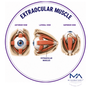
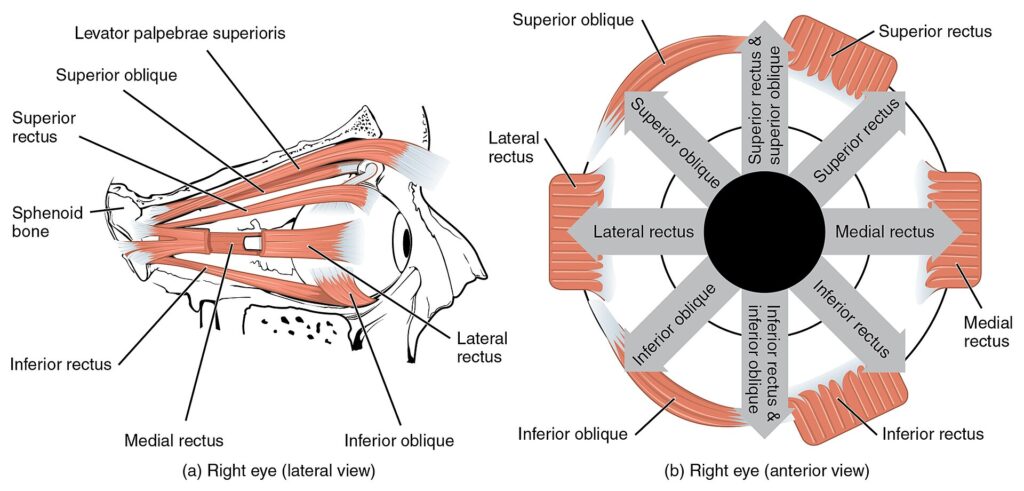
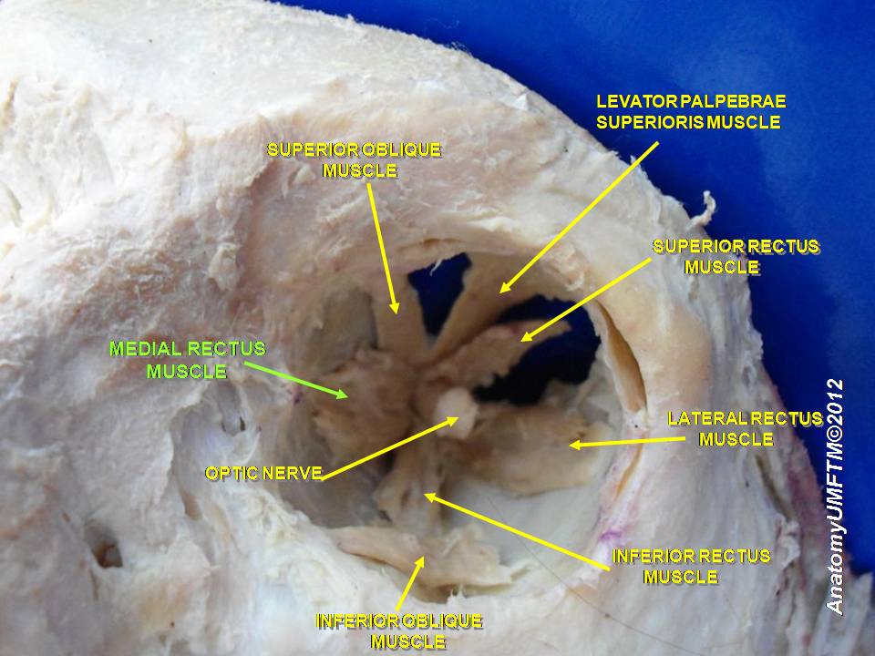
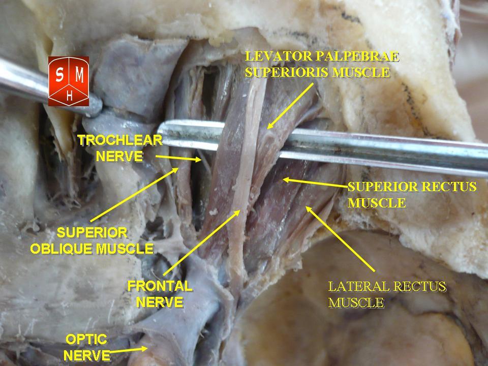
Extraocular Muscle
MYO CORE
Levator Palpebrae Superioris
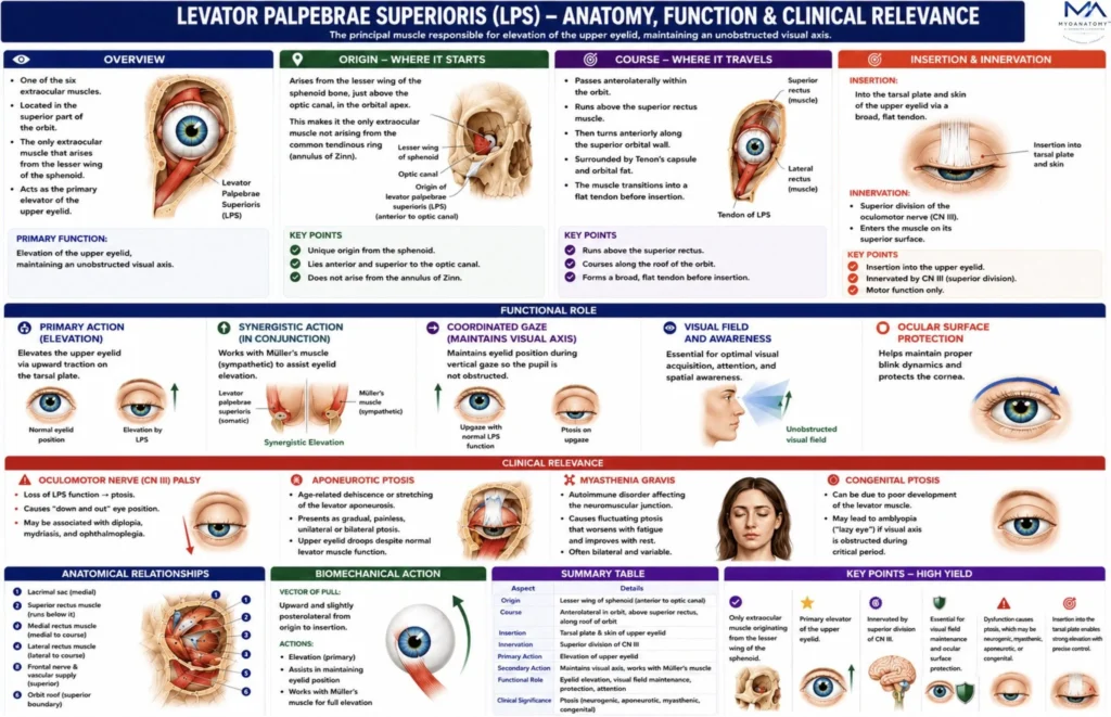
The levator palpebrae superioris (LPS) is the principal skeletal muscle responsible for elevation of the upper eyelid, thereby maintaining an unobstructed visual axis. Although it does not directly act on the eyeball itself, it forms an integral component of the orbital motor apparatus, functioning in coordinated association with the extraocular muscles – particularly the superior rectus muscle – to synchronize eyelid position with vertical gaze.
OVERVIEW
The muscle originates from the lesser wing of the sphenoid bone at the orbital apex, superior to the optic canal, and extends anteriorly along the superior orbital wall.
Anteriorly, the muscular belly transitions into a broad fibrous expansion known as the levator aponeurosis, which inserts into the superior tarsal plate and the dermis of the upper eyelid, thereby transmitting muscular force to the eyelid.
Through its interaction with Whitnall’s ligament, the superior tarsal muscle, and the orbital fascial system, it forms a highly specialized mechanism that ensures precise eyelid positioning during ocular movement. Its activity is regulated by oculomotor and sympathetic pathways, and dysfunction results in ptosis, a key diagnostic sign in neurological and ophthalmological disorders.
Functionally, eyelid elevation is achieved through a dual system:
Voluntary skeletal component – levator palpebrae superioris (innervated by CN III)
Autonomic tonic component – superior tarsal muscle (Müller’s muscle), innervated by sympathetic fibers
This dual mechanism ensures both active eyelid elevation and maintenance of tonic eyelid position, preventing eyelid drooping during wakefulness.


ANATOMY
Origin
The levator palpebrae superioris arises from the:
inferior surface of the lesser wing of the sphenoid bone, immediately superior to the optic canal and anterior to the annulus of Zinn.
This origin lies:
superior to the origin of the superior rectus muscle
medial to the lacrimal gland fossa
The muscle belly therefore occupies the superior extraconal compartment of the orbit.
Exam Question
Evaluate the anatomical importance of the levator palpebrae superioris origin from the lesser wing of the sphenoid. Explain how its relationship to the optic canal, annulus of Zinn, and superior rectus muscle influences orbital organization and surgical anatomy.
Course
From its origin, the levator muscle runs anteriorly along the roof of the orbit, positioned between:
the orbital roof (frontal bone) superiorly
the superior rectus muscle inferiorly.
The muscle belly remains muscular for approximately the posterior two-thirds of its course.
In the anterior orbit, the muscle fibers transition into the levator aponeurosis, a broad fibrous sheet that continues forward toward the eyelid.
Exam Question
Discuss how the superior orbital course of the levator palpebrae superioris positions it within the superior extraconal compartment. Analyze the functional significance of its relationship to the orbital roof and superior rectus during ocular movement.
Insertion
The levator aponeurosis represents the terminal tendon of the levator palpebrae superioris. It expands anteriorly and divides into several attachments:
Primary insertions
anterior surface of the superior tarsal plate
dermis of the upper eyelid
These dermal insertions form the upper eyelid crease, which is visible externally.
Secondary attachments
Fibers also extend to: the orbital septum surrounding connective tissue of the eyelid, fibers of orbicularis oculi
These attachments stabilize eyelid motion during blinking and eye movements.
Superior Tarsal Muscle (Müller’s Muscle) – levator aponeurosis gives rise to a thin sheet of smooth muscle fibers, known as the superior tarsal muscle (Müller’s muscle).
origin – undersurface of the levator aponeurosis.
insertion- superior border of the superior tarsal plate.
Innervation – Sympathetic fibers from the superior cervical ganglion
Function Provides tonic elevation of the eyelid, maintaining eyelid opening during alertness.
Loss of sympathetic tone leads to partial ptosis.
Fascial and Connective Tissue System
The levator muscle is integrated into the orbital fascial framework, which coordinates eyelid motion with ocular movement.

Exam Question
Analyze the biomechanical significance of the levator aponeurosis. Explain how insertion into the superior tarsal plate, dermis of the upper eyelid, orbital septum, and orbicularis oculi contributes to coordinated eyelid elevation and formation of the upper eyelid crease.
Important Structures
Tenon’s Capsule (Fascia Bulbi)-
a fibrous sheath surrounding the globe and extraocular muscles, providing a gliding surface for ocular movement.
Orbital Septum
a fibrous membrane extending from the orbital rim to the tarsal plates, separating orbital contents from eyelid tissues.
Whitnall’s Ligament (Superior Transverse Ligament)
a condensation of connective tissue that acts as a pulley for the levator muscle, changing its direction from horizontal to vertical as it approaches the eyelid. Ligament therefore:
stabilizes the levator muscle; prevents posterior displacement of the globe during contraction.
Exam Question
Critically evaluate the functional roles of Whitnall’s ligament, Tenon’s capsule, the orbital septum, and Müller’s muscle in supporting levator palpebrae superioris activity. Explain how disruption of these structures may alter eyelid mechanics.
Innervation
The levator palpebrae superioris is innervated by the superior division of CN III, while
sympathetic fibers supply Müller’s muscle.
bilateral control arises from the central caudal nucleus of the midbrain, ensuring synchronous eyelid elevation.
During upward gaze, the levator acts in coordination with the superior rectus to maintain an unobstructed visual field.
Dysfunction may result in ptosis, lid lag, or impaired eyelid movement
Exam Question
Describe the neural control of the levator palpebrae superioris. Analyze the respective contributions of the superior division of the oculomotor nerve, the central caudal nucleus, and sympathetic fibers to normal eyelid position and movement.
FUNCTIONAL ROLE
Visual Maintainence
The levator palpebrae superioris is the principal elevator of the upper eyelid, maintaining a patent visual axis and ensuring uninterrupted exposure of the pupil.
Continuous activity of the muscle is essential for normal visual acquisition, spatial awareness, and effective interaction with the external environment
Exam Question
Why is continuous activity of the levator palpebrae superioris essential for normal visual acquisition? Discuss how eyelid elevation contributes to visual-field preservation, environmental awareness, and ocular function
Corneal Protection
By sustaining appropriate eyelid position, the levator prevents mechanical encroachment upon the cornea and preserves the functional integrity of the ocular surface.
This balance allows adequate corneal exposure for vision while supporting normal tear-film distribution and ocular surface homeostasis
Exam Question
Explain how appropriate eyelid positioning by the levator palpebrae superioris contributes to corneal exposure, tear-film stability, and maintenance of ocular surface homeostasis.
Gaze Synchronization
The levator operates as part of an integrated oculopalpebral system, dynamically coordinating eyelid movement with globe position.
During ocular excursions, particularly vertical gaze, this synchronization ensures that the upper eyelid does not obstruct the visual field and maintains optimal visual performance.
Exam Question
Analyze the mechanisms by which levator palpebrae superioris activity is synchronized with globe movement. Discuss the importance of coordinated eyelid and ocular motion during vertical gaze.
Alertness Regulation
The muscle exhibits continuous tonic contraction throughout wakefulness and is closely linked to central neural and sympathetic pathways governing arousal.
Consequently, eyelid position serves as an important indicator of neurological alertness, attentional state, and overall level of consciousness
Exam Question
Evaluate the relationship between levator palpebrae superioris tone, sympathetic activity, and neurological arousal. Explain why eyelid position is considered an important clinical indicator of consciousness and alertness
CLINICAL RELEVANCE
Oculomotor Palsy
Lesions of the oculomotor nerve (CN III) abolish motor input to the levator palpebrae superioris, producing complete ptosis.
Because CN III also innervates most extraocular muscles and carries parasympathetic fibers, affected patients typically exhibit a down-and-out eye position, diplopia, and pupillary dilation.
Exam Question
Analyze the pathophysiological mechanisms by which oculomotor nerve (CN III) lesions produce complete ptosis. Correlate levator paralysis with associated findings including ophthalmoplegia, pupillary dilation, and the characteristic “down-and-out” eye position
Horner Syndrome
Disruption of the sympathetic pathway to Müller’s muscle results in partial ptosis, reflecting loss of tonic eyelid elevation rather than complete levator paralysis.
This classic syndrome is commonly associated with miosis and anhidrosis, indicating sympathetic dysfunction
Exam Question
Evaluate the anatomical and physiological basis of partial ptosis in Horner syndrome. Compare sympathetic denervation of Müller’s muscle with complete levator palpebrae superioris paralysis caused by oculomotor nerve injury.
Myasthenis Gravis
Myasthenia gravis frequently involves the levator palpebrae superioris due to autoimmune impairment of neuromuscular transmission.
The resulting weakness produces fluctuating ptosis, diplopia, and fatigability, with symptoms typically worsening during sustained eyelid elevation and improving with rest.
Exam Question
Discuss why the levator palpebrae superioris is frequently affected in myasthenia gravis. Analyze the mechanisms responsible for fluctuating ptosis and explain how neuromuscular junction dysfunction alters eyelid elevation
Aponeurotic Ptosis
Age-related stretching, attenuation, or dehiscence of the levator aponeurosis reduces efficient transmission of levator force to the upper eyelid.
This produces a characteristic drooping eyelid despite preserved muscle function and represents the most common cause of involutional ptosis.
Exam Question
Critically evaluate the development of aponeurotic ptosis. Explain how age-related attenuation, stretching, or dehiscence of the levator aponeurosis results in eyelid drooping despite preserved muscle function and neural innervation.
SUMMARY TABLE