Suprahyoid Muscle

MYO CORE

Digastric Muscle

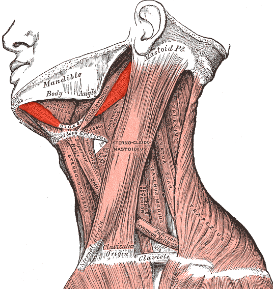
The digastric muscle is a paired suprahyoid muscle located in the anterior region of the neck, extending between the mandible and the hyoid bone. It plays an important role in mandibular depression (opening of the mouth), stabilization of the hyoid bone, and swallowing mechanics

OVERVIEW

The name digastric (Greek di = two, gaster = belly) reflects its characteristic structure: the muscle consists of two muscular bellies connected by an intermediate tendon. These bellies have different embryological origins and nerve supplies, which is unique among muscles of the head and neck.

The digastric muscle forms an important anatomical landmark in the submandibular region, contributing to the boundaries of the submandibular triangle and submental triangle. Through these relationships it plays a key role in surgical anatomy of the neck, particularly during procedures involving the submandibular gland, facial vessels, and carotid region.

Functionally, the digastric muscle acts as a link between mandibular movement and hyoid movement, integrating the mechanics of mastication, swallowing, and speech

ANATOMY

Origin

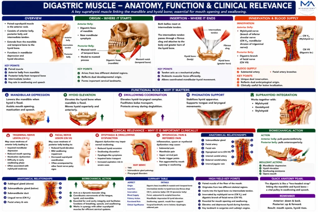
Anterior Belly – originates from – digastric fossa of the mandible (on the inner surface of the mandibular body near the midline)

Posterior Belly – originates from – mastoid notch of the temporal bone (medial to the mastoid process)

Exam Question

Evaluate the embryological and anatomical significance of the dual origin of the digastric muscle and discuss how these origins influence its morphology and function.

Insertion

The two bellies meet at the intermediate tendon, which is connected to: the body of the hyoid bone via a fibrous loop attached to the greater horn of the hyoid.

This fibrous sling allows the tendon to slide during contraction while maintaining its attachment to the hyoid bone.

Exam Question

Explain the biomechanical importance of the intermediate tendon and fibrous hyoid sling, and analyze how this arrangement optimizes force transmission during swallowing and mandibular depression.

Anatomical Structure

The digastric muscle consists of two distinct portions:

Anterior Belly- located in the submental region derived from the first pharyngeal arch

Posterior Belly-located in the upper lateral neck derived from the second pharyngeal arch.

These two bellies are connected by the intermediate tendon, which is attached to the hyoid bone by a fibrous sling.

This arrangement allows the muscle to change the direction of force during contraction, enabling coordinated movement between the mandible and the hyoid apparatus.

Exam Question

Compare the anterior and posterior bellies of the digastric muscle with respect to location, embryological derivation, morphology, and functional specialization.

Innervation

The digastric muscle has dual innervation, reflecting its dual embryological origin.

Anterior Belly -innervated by: – mylohyoid nerve, branch of:  inferior alveolar nerve (mandibular division of trigeminal nerve — CN V₃)

Posterior Belly -innervated by:  digastric branch of the facial nerve (CN VII)

This dual innervation is an important anatomical feature used in neuroanatomical localization of nerve lesions.

Exam Question

Analyze the clinical and embryological significance of the dual innervation of the digastric muscle and explain how this feature aids neuroanatomical lesion localization.

FUNCTIONAL ROLE

Mandibular Depression

The digastric muscle serves as an important depressor of the mandible when the hyoid bone is stabilized by the infrahyoid musculature. 

Contraction lowers the mandible and contributes directly to mouth opening, facilitating essential functions such as mastication, speech articulation, and the preparatory phases of swallowing. Through this action, the muscle forms a functional link between the suprahyoid region and the masticatory apparatus

Exam Question

Evaluate the role of the digastric muscle in mandibular depression and discuss its contribution to mastication, speech articulation, and mouth opening.

Hyoid Elevation

When the mandible remains fixed, contraction of the digastric muscle elevates the hyoid bone in a superior and anterior direction. 

This movement is a critical component of the early swallowing mechanism, helping to reposition the hyoid-laryngeal complex and facilitate safe passage of the food bolus through the pharynx. Effective hyoid elevation also contributes to airway protection during deglutition.

Exam Question

Analyze the biomechanical role of the digastric muscle in hyoid elevation during deglutition and explain its importance in airway protection and bolus transport.

Functional Integration

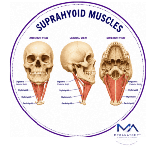
The digastric muscle operates as part of the coordinated suprahyoid muscular complex together with the mylohyoid, geniohyoid, and stylohyoid muscles. Collectively, these muscles regulate mandibular movement, hyoid elevation, tongue stabilization, and swallowing biomechanics. 

By stabilizing the hyoid apparatus, which serves as an anchoring platform for the tongue, pharyngeal, and laryngeal muscles, the digastric muscle plays a central role in coordinated phonation and deglutition

Exam Question

Discuss how the digastric muscle functionally integrates with the mylohyoid, geniohyoid, and stylohyoid muscles to coordinate swallowing, phonation, and mandibular movement.

Anatomical Relation

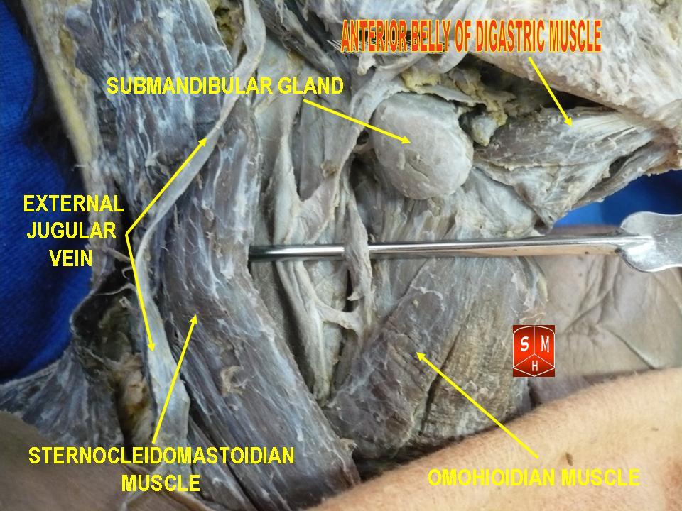
The digastric muscle is a major anatomical landmark of the upper neck. Its anterior and posterior bellies form the boundaries of the submandibular triangle, which contains the submandibular gland, facial vessels, and regional lymph nodes. 

The paired anterior bellies also define the submental triangle, housing submental lymph nodes and tributaries of the anterior jugular vein. Posteriorly, the muscle is closely related to the internal and external carotid arteries, internal jugular vein, and hypoglossal nerve (CN XII), making it an important surgical landmark during cervical and vascular procedures

Exam Question

Evaluate the importance of the digastric muscle as a surgical landmark and explain its relationships to the submandibular triangle, carotid region, and major neurovascular structures.

CLINICAL RELEVANCE

Surgical Importance

The digastric muscle serves as a critical surgical landmark within the upper neck. 

Its bellies assist in identifying major structures including the carotid arteries, hypoglossal nerve (CN XII), facial artery, and submandibular gland, making it particularly valuable during cervical and submandibular procedures

Exam Question

Analyze the surgical relevance of the digastric muscle during cervical and submandibular procedures and discuss how it assists in identifying critical anatomical structures.

Neural Injury

Neurological lesions may impair digastric function depending on the affected nerve. 

Facial nerve (CN VII) injury can paralyze the posterior belly, compromising hyoid stabilization and swallowing efficiency, whereas mandibular nerve (V₃) lesions affect the anterior belly, potentially impairing mandibular depression and masticatory coordination.

Exam Question

Compare the functional consequences of trigeminal nerve (CN V₃) and facial nerve (CN VII) lesions on digastric muscle function and related head and neck movements.

Operative Guidance

During submandibular gland surgery, the digastric muscle provides an important anatomical guide for safe dissection. 

Its predictable relationships help surgeons identify and preserve adjacent neurovascular structures, reducing the risk of operative complications

Exam Question

Explain how knowledge of digastric muscle anatomy improves operative safety during submandibular gland surgery and cervical dissections.

Referd Pain

Inflammation, spasm, or myofascial dysfunction of the digastric muscle may produce referred pain within the mandibular, submental, or upper cervical regions. 

Symptoms can mimic temporomandibular disorders, odontogenic pain, or pharyngeal discomfort, creating diagnostic challenges in head and neck practice

Exam Question

Evaluate the mechanisms by which digastric muscle dysfunction may produce referred pain and discuss its differential diagnostic significance in head and neck disorders.

SUMMARY TABLE

Scroll to Top