Suprahyoid Muscle

MYO CORE

Mylohyoid Muscle

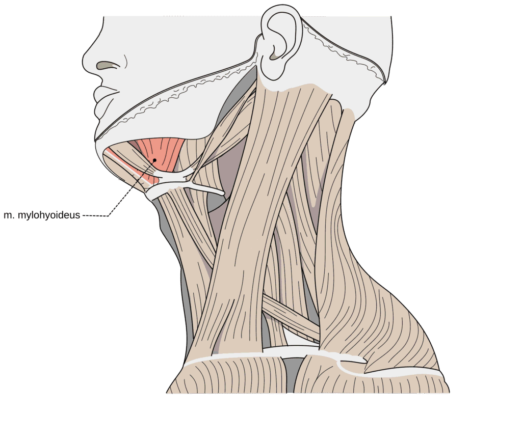
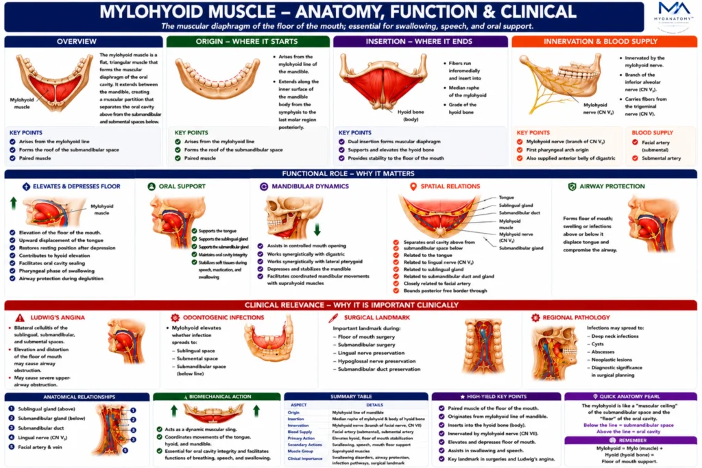
The mylohyoid muscle is a flat, triangular suprahyoid muscle that forms the muscular floor of the oral cavity. It extends between the mandible and the hyoid bone, creating a muscular diaphragm that separates the oral cavity above from the submandibular and submental spaces below.

OVERVIEW

Functionally, the mylohyoid plays a central role in swallowing, speech, and mandibular movement, acting as a dynamic support structure for the tongue and hyoid apparatus. Because of its anatomical position, it contributes to the mechanics of mastication, deglutition, and oral stabilization.

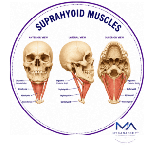
The muscle works closely with the digastric, geniohyoid, and stylohyoid muscles, forming the suprahyoid complex, which coordinates movements between the mandible, hyoid bone, and tongue.

In clinical anatomy, the mylohyoid muscle is highly significant because it forms an important anatomical barrier that influences the spread of odontogenic infections between the sublingual and submandibular spaces.

ANATOMY

Origin

Originates from themylohyoid line of the mandible

This ridge runs along the inner surface of the mandibular body, extending from the mandibular symphysis anteriorly to the last molar region posteriorly.

Exam Question

How does the origin of the mylohyoid from the mylohyoid line support floor-of-mouth stability and mandibulohyoid movement?

Insertion

The muscle fibers run inferomedially and insert into two structures:

median raphe of the mylohyoid muscle

body of the hyoid bone

The mylohyoid raphe is a fibrous midline structure where the left and right mylohyoid muscles meet.

Exam Question

Why is the dual insertion of the mylohyoid into the median raphe and hyoid bone essential for forming the muscular diaphragm of the oral cavity?

Innervation

The mylohyoid muscle is innervated by the – mylohyoid nerve branch of  inferior alveolar nerve  which arises from the mandibular division of the trigeminal nerve (CN V₃)

This nerve also supplies the anterior belly of the digastric muscle, reflecting their common first pharyngeal arch origin.

Exam Question

How does first pharyngeal arch development explain the innervation of the mylohyoid by the mylohyoid nerve (CN V₃)?

FUNCTIONAL ROLE

Swallowing Function

The mylohyoid is a principal muscle of the oral and early pharyngeal phases of swallowing. By elevating the floor of the mouth and supporting upward displacement of the tongue, it facilitates posterior propulsion of the food bolus toward the pharynx. 

Simultaneous elevation of the hyoid bone contributes to laryngeal ascent, enhancing airway protection and ensuring efficient coordination of deglutition.

Exam Question

Explain the role of the mylohyoid in bolus propulsion, hyoid elevation, and airway protection during swallowing.

Oral Support

Acting as the muscular diaphragm of the floor of the mouth, the mylohyoid provides structural support for the tongue, sublingual gland, and submandibular duct.

It maintains the anatomical integrity of the oral cavity while stabilizing soft tissues during speech, mastication, swallowing, and complex lingual movements. This supportive role is essential for preserving normal oral function and biomechanical stability.

Exam Question

How does the mylohyoid provide structural support for the tongue and floor of the mouth during speech and mastication?

Mandibular Dynamics

When the hyoid bone is stabilized by the infrahyoid musculature, the mylohyoid assists in mandibular depression and controlled mouth opening. 

Working synergistically with the digastric and lateral pterygoid muscles, it contributes to coordinated mandibular movement required for speech, mastication, respiration, and oral manipulation of food. Its dual attachment to both mandible and hyoid enables integration of oral and cervical biomechanics.

Exam Question

How does the mylohyoid assist mandibular depression and controlled mouth opening when the hyoid bone is stabilized?

Spatial Relation

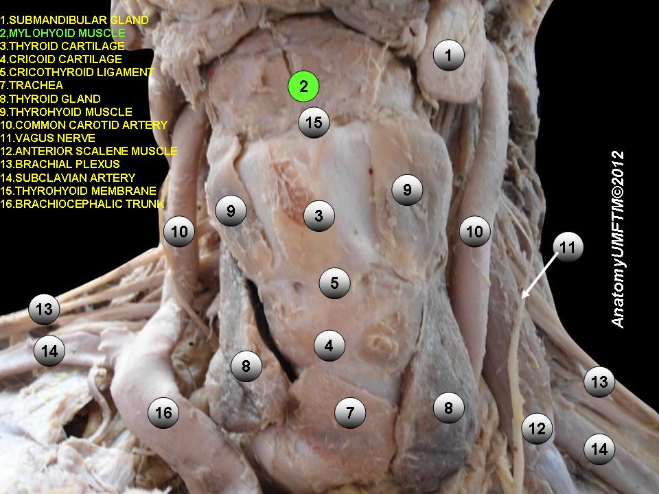
The mylohyoid forms the anatomical boundary between the sublingual and submandibular compartments. Superiorly, it is related to the tongue, lingual nerve, sublingual gland, and submandibular duct, whereas inferiorly it is associated with the submandibular gland, facial vessels, and regional lymphatics. 

Its free posterior border permits communication between the sublingual and submandibular spaces, creating an important pathway for the spread of odontogenic infections and deep neck space pathology.

Exam Question

Why is the mylohyoid considered the principal anatomical boundary between the sublingual and submandibular spaces?

CLINICAL RELEVANCE

Infection Pathways

The mylohyoid is a critical anatomical barrier that governs the spread of odontogenic infections arising from the mandibular dentition. Infections originating superior to the mylohyoid line typically extend into the sublingual space, whereas those arising inferior to the line preferentially involve the submandibular space. 

This relationship is fundamental to the anatomical localization, radiological interpretation, and clinical management of deep neck infections.

Exam Question

How does the position of the mylohyoid determine the spread of odontogenic infections into the sublingual or submandibular spaces?

Airway Protection

The anatomical position of the mylohyoid is central to the pathogenesis of Ludwig’s angina, a rapidly progressive cellulitis involving the sublingual and submandibular compartments. 

Inflammatory edema within these spaces elevates the floor of the mouth and displaces the tongue posteriorly, creating a potentially life-threatening risk of upper airway obstruction. Detailed understanding of mylohyoid anatomy is therefore essential for timely diagnosis and airway management

Exam Question

Why is understanding mylohyoid anatomy critical in the development and management of Ludwig’s angina?

Surgical Landmark

The mylohyoid serves as a major surgical landmark during procedures involving the floor of the mouth and submandibular region. Its position assists in identifying and protecting critical structures including the submandibular duct, lingual nerve, and hypoglossal nerve.

Accurate recognition of these relationships minimizes operative complications and preserves salivary, sensory, and motor function.

Exam Question

Why is the mylohyoid an important surgical landmark during procedures involving the floor of the mouth and submandibular region?

Regional Pathology

Because the mylohyoid separates the sublingual and submandibular spaces while permitting communication through its posterior free border, pathological processes may spread across fascial compartments of the floor of the mouth.

This anatomical arrangement influences the progression of infections, cysts, abscesses, and neoplastic lesions, making the muscle an important structure in both diagnostic evaluation and surgical planning.

Exam Question

How does the anatomical position of the mylohyoid influence the spread of infections and other pathologies within the floor of the mouth?

SUMMARY TABLE

Scroll to Top