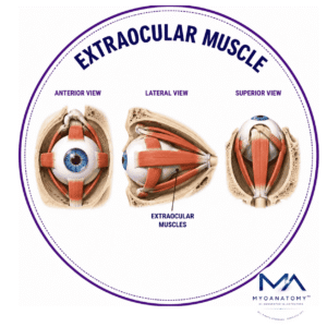
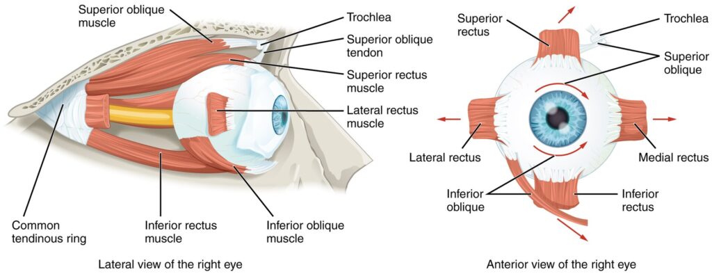
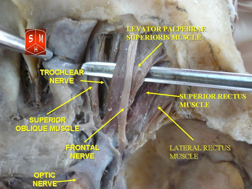
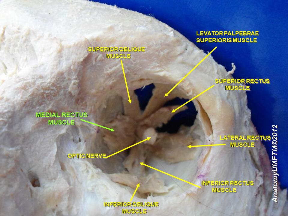
Extraocular Muscle

MYO CORE

Superior Rectus Muscle

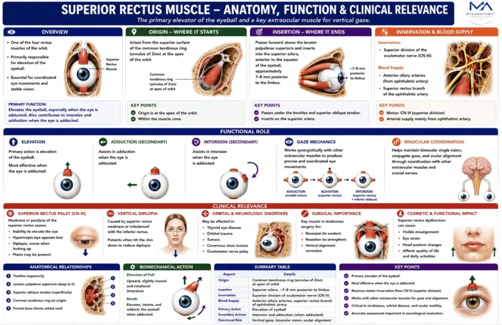
The superior rectus is one of the four rectus muscles of the orbit and one of the principal extraocular muscles responsible for controlling movement of the eyeball. It extends from the orbital apex to the superior anterior sclera, lying immediately beneath the roof of the orbit and superior to the globe

OVERVIEW

Functionally, it is primarily an elevator of the eye, but because the orbital axis and visual axis do not coincide, its action is more complex than simple upward movement. In addition to elevation, it also contributes to adduction and intorsion.

The muscle is part of the highly specialized neuromuscular system that allows precise ocular alignment, conjugate gaze, fixation, and binocular vision. Its action must be interpreted in relation to the primary position of gaze, the orientation of the orbit, and the coordinated activity of the other extraocular muscles.

ANATOMY

Origin

The superior rectus originates from the:

common tendinous ring (annulus tendineus communis, annulus of Zinn) at the apex of the orbit

This fibrous ring surrounds:  the optic canal ;  part of the superior orbital fissure

The common tendinous ring serves as the shared origin for the four rectus muscles:

superior rectus; inferior rectus

medial rectus; lateral rectus

From this origin, the superior rectus passes anteriorly along the superior aspect of the orbit

Exam Question

Explain the anatomical significance of the common tendinous ring (annulus of Zinn) as the origin of the superior rectus muscle and discuss how this arrangement contributes to coordinated extraocular muscle function

Insertion

The superior rectus inserts into the:

superior surface of the sclera, anterior to the equator of the eyeball

More precisely, it inserts into the sclera approximately 7–8 mm posterior to the corneoscleral junction (limbus). Its insertion is broad and curved, conforming to the contour of the globe.

Like the other rectus muscles, it inserts anterior to the equator of the eyeball, which is important for understanding its vector of action.

Exam Question

Describe the insertion of the superior rectus muscle on the sclera and analyze how the position of its attachment relative to the equator of the globe influences its primary and secondary actions.

Anatomical Relation

The superior rectus has important relations within the superior orbital compartment.

Superior relations- levator palpebrae superioris, which lies above it

orbital roof (mainly frontal bone)

Inferior relation  – superior surface of the eyeball

Medial relations – superior oblique tendon region (anteriorly, more medially)

Lateral relations – lacrimal nerve and superior ophthalmic vessels in the superior orbit

Because the levator palpebrae superioris lies directly superior to it, the superior rectus is closely related to eyelid mechanics as well as globe movement.

Fiber Direction and Mechanical Orientation

Although called a “rectus” muscle, the superior rectus does not act purely in a vertical plane. This is because:

the orbital axis is directed anterolaterally

the visual axis is directed straight anteriorly

The angle between these axes is approximately 23 degrees.

As a result, contraction of the superior rectus produces a combined vector with three components:

Elevation; Adduction; Intorsion

Thus, the superior rectus is not a pure elevator in all positions of gaze.

Exam Question

Discuss the anatomical relations of the superior rectus muscle within the superior orbital compartment and explain the functional importance of its relationship to the levator palpebrae superioris and orbital roof.

Innervation

The superior rectus is innervated by the:

superior division of the oculomotor nerve (cranial nerve III)

The oculomotor nerve enters the orbit through the superior orbital fissure and divides into superior and inferior divisions.

The superior division supplies:

superior rectus

levator palpebrae superioris

This close shared innervation explains why lesions of the superior division of CN III may affect both eyeball elevation and upper eyelid elevation.

Exam Question

Describe the motor innervation of the superior rectus muscle, including its cranial nerve supply and orbital course, and explain the clinical consequences of interruption of this neural pathway.

FUNCTIONAL ROLE

Ocular Elevation

The superior rectus serves as the principal elevator of the eyeball, generating upward movement of the visual axis. 

It is the primary muscle responsible for vertical gaze above the horizontal plane and plays a critical role in coordinated upward ocular movements

Exam Question

How do the anatomical orientation and line of pull of the superior rectus muscle enable elevation of the globe, and why is its action influenced by eye position within the orbit?

Rotational Control

In addition to elevation, the superior rectus contributes to adduction and intorsion of the globe. These secondary actions stabilize ocular orientation, maintain retinal alignment, and assist in preserving accurate visual tracking during complex eye movements

Exam Question

Explain the biomechanical basis of the superior rectus muscle’s secondary actions (adduction and intorsion) and discuss their importance in maintaining ocular stability during complex gaze movements.

Gaze Mechanics

The functional effect of the superior rectus varies according to eye position. 

When the eye is abducted, it acts as a nearly pure elevator; in primary gaze it produces elevation, adduction, and intorsion simultaneously, demonstrating the biomechanical complexity of extraocular muscle action.

Exam Question

Analyze how the functional contribution of the superior rectus muscle changes during primary gaze, abduction, and adduction, and explain the clinical significance of these variations during ocular examination.

Binocular Coordination

The superior rectus functions within integrated ocular motor networks to maintain binocular single vision, conjugate upward gaze, globe stabilization, and precise visual fixation. 

Through coordination with fellow extraocular muscles, cranial nerve pathways, vestibulo-ocular reflex circuits, and brainstem gaze centers, it ensures stable, diplopia-free vision during head and eye movements.

Exam Question

Evaluate the role of the superior rectus muscle in binocular vision and conjugate gaze, emphasizing its integration with brainstem gaze centers and vestibulo-ocular reflex pathways.

CLINICAL RELEVANCE

Oculomotor Palsy

Lesions of the oculomotor nerve (CN III) may impair or paralyze the superior rectus, resulting in reduced upward gaze, vertical diplopia, and contributing to the characteristic “down-and-out” eye position of complete oculomotor nerve palsy. 

Associated ptosis is common due to concurrent involvement of the levator palpebrae superioris

Exam Question

Explain the pathophysiological mechanisms by which oculomotor nerve (CN III) lesions produce superior rectus dysfunction, and discuss the resulting ocular posture, motility deficits, and visual symptoms.

Vertical Diplopia

Superior rectus weakness produces impaired ocular elevation and vertical ocular misalignment, leading to diplopia that becomes more pronounced during upward gaze. 

Patients frequently adopt compensatory head postures to maintain binocular single vision and reduce visual discomfort.

Exam Question

Evaluate the anatomical and functional basis of vertical diplopia resulting from superior rectus weakness and explain the compensatory mechanisms adopted by affected patients.

Orbital Pathology

Restrictive disorders such as thyroid eye disease, orbital trauma, and postoperative fibrosis may mechanically limit superior rectus function. 

These conditions disrupt normal globe mobility and commonly present with restricted upward gaze and abnormal ocular motility patterns.

Exam Question

Explain how orbital disorders such as thyroid eye disease, trauma, and postoperative fibrosis may impair superior rectus function, and discuss their effects on ocular motility and gaze mechanics.

Strabismus Correction

The superior rectus is a key structure in strabismus surgery, orbital decompression procedures, and superior orbital approaches. 

Functional imbalance between the superior rectus and its antagonists or synergists may result in vertical strabismus, hypertropia, hypotropia, and gaze-dependent ocular misalignment, making the muscle particularly important in pediatric ophthalmology and neuro-ophthalmology.

Exam Question

Evaluate the role of the superior rectus muscle in the pathogenesis and surgical correction of vertical strabismus, including the principles underlying superior rectus recession and resection procedures.

SUMMARY TABLE

Scroll to Top