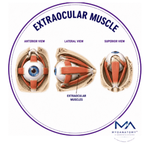
Extraocular Muscle
MYO CORE
Inferior Oblique Muscle
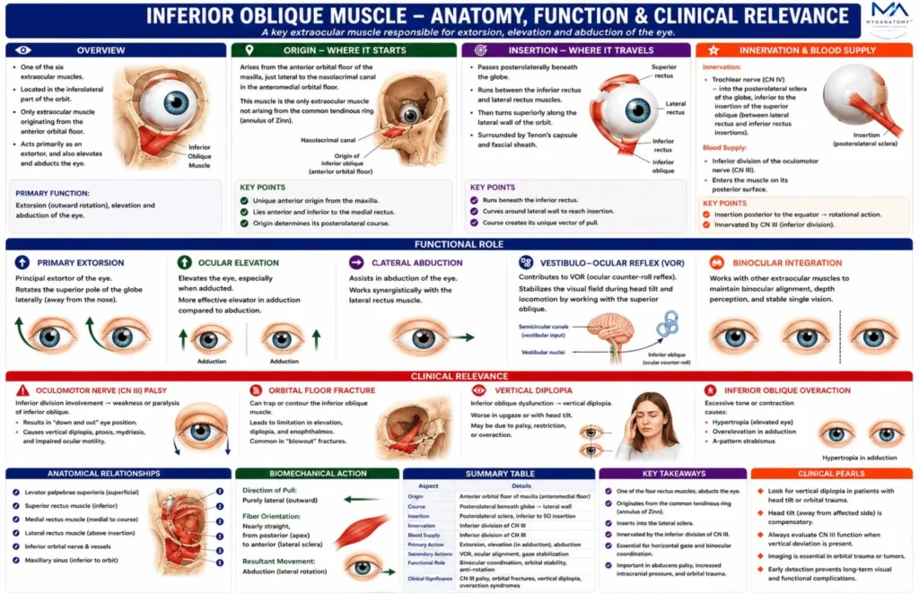
The inferior oblique muscle is one of the six extraocular muscles responsible for controlling ocular orientation and torsional stability within the orbit. It belongs to the oblique group of extraocular muscles, together with the superior oblique, and plays a fundamental role in ocular torsion, vertical gaze coordination, and stabilization of the visual field during head movement
OVERVIEW
The inferior oblique muscle is a unique extraocular muscle originating from the anterior orbital floor of the maxilla and inserting on the posterolateral sclera of the eyeball. Its contraction produces extorsion, elevation, and abduction, with extorsion representing its primary action.
Through its interaction with the superior oblique and vertical rectus muscles, the inferior oblique plays a crucial role in torsional balance of the eyeball, stabilization of the visual field during head movement, and coordinated vertical gaze.
Dysfunction of this muscle may result in vertical diplopia, abnormal ocular torsion, and strabismus, making it clinically important in neurological and ophthalmological examinations.
The muscle receives motor innervation from the inferior division of the oculomotor nerve (cranial nerve III) and participates in coordinated ocular movement systems involving the vestibulo-ocular reflex (VOR), vertical gaze centers of the midbrain, and binocular visual alignment mechanisms.
ANATOMY
Origin
Originates from the:
orbital surface of the maxilla, specifically the anteromedial orbital floor just lateral to the nasolacrimal canal.
This origin lies:
posterior to the lacrimal sac
lateral to the nasolacrimal duct
inferior to the medial orbital wall
This position makes the inferior oblique the only extraocular muscle originating from the anterior orbit rather than the orbital apex.
Because of this anterior origin, the inferior oblique acts from an anterior-to-posterior direction, unlike the rectus muscles which act from posterior to anterior.
Exam Question
Analyze the anatomical significance of the inferior oblique being the only extraocular muscle that originates from the anterior orbital floor rather than the common tendinous ring, and explain how this origin determines its functional vector of pull.
Course
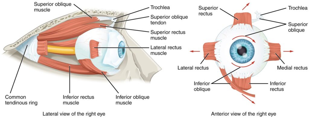
From its origin, the muscle travels: posteriorly laterally superiorly
The muscle runs beneath the inferior rectus muscle, crossing the inferior surface of the globe within the extraconal orbital fat compartment.
As it approaches the posterolateral aspect of the eyeball, the muscle transitions into a short tendon, which then attaches to the sclera.
The oblique trajectory across the globe generates a rotational torque, which is responsible for the muscle’s torsional action.
Exam Question
Discuss how the inferolateral and posterior course of the inferior oblique beneath the globe contributes to its combined extorsional, elevating, and abducting actions. Relate your answer to ocular rotational mechanics.
Anatomical Relations
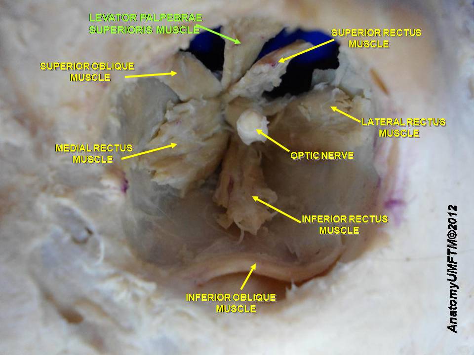
Superior – inferior rectus muscle
Medial – nasolacrimal duct; Lacrimal sac
Inferior – orbital floor (maxillary bone)
Lateral – lateral rectus muscle
Posterior – posterolateral sclera
Neurovascular relations
infraorbital nerve and artery
branches of the ophthalmic artery.
Biomechanics of Inferior Oblique Contraction
Because the muscle attaches posterior to the equator of the globe and runs obliquely across its inferior surface, contraction produces a three-component rotational movement
Exam Question
Evaluate the anatomical relationships of the inferior oblique muscle with the lacrimal apparatus, orbital floor, inferior rectus, and neurovascular structures, and discuss their importance during orbital and strabismus surgery.
Fascial System
The inferior oblique is enclosed by a connective tissue sheath continuous with Tenon’s capsule (fascia bulbi).
Several fascial structures stabilize the muscle.
Inferior Oblique Fascial Sheath- blends with orbital connective tissue and provides mechanical stabilization during contraction.
Lockwood’s Suspensory Ligament
The inferior rectus and inferior oblique contribute fibers to Lockwood’s ligament, a hammock-like structure supporting the globe.
This structure:
stabilizes the eyeball within the orbit; prevents downward displacement of the globe.
Exam Question
Analyze the contribution of Tenon’s capsule, the inferior oblique fascial sheath, and Lockwood’s suspensory ligament to orbital biomechanics and globe stabilization. Explain the consequences of disruption of these structures.
Orbital System
Modern orbital biomechanics demonstrates that extraocular muscles function through fibroelastic pulleys located within orbital connective tissue.
The inferior oblique interacts with these pulleys to maintain accurate muscle vectors during eye movement.
Disruption of pulley alignment can result in strabismus or abnormal torsion.
Exam Question
Discuss the role of orbital connective-tissue pulleys in regulating inferior oblique muscle function and evaluate how pulley dysfunction may contribute to complex strabismus syndromes.
Insertion
Inserts into the:
posterolateral sclera of the eyeball, inferior to the insertion of the superior oblique.
Important characteristics of the insertion:
located posterior to the equator of the globe
lies between the lateral rectus and inferior rectus muscles
spreads across a relatively broad scleral area
Because the insertion lies posterior to the globe’s equator, contraction produces rotational movement rather than simple linear translation.
Exam Question
Explain why insertion of the inferior oblique posterior to the equator of the globe permits rotational ocular movement rather than simple translational displacement. Correlate the insertion site with the muscle’s primary and secondary actions.
Innervation
The inferior oblique receives motor innervation from the:
Inferior division of the oculomotor nerve (cranial nerve III).
The inferior branch of CN III supplies:
inferior rectus; medial rectus
inferior oblique
The nerve enters the muscle on its posterior surface, ensuring efficient transmission of motor impulses.
Exam Question
Describe the motor innervation of the inferior oblique muscle by the inferior division of the oculomotor nerve and analyze the clinical manifestations that may arise following selective injury to this neural pathway.
FUNCTIONAL ROLE
Primary Extorsion
The inferior oblique is the principal extortor of the eyeball. Contraction rotates the superior pole of the globe laterally away from the nasal side, producing external ocular torsion.
This rotational movement is essential for maintaining visual orientation during head tilt and contributes significantly to stabilization of the retinal image within the visual field
Exam Question
Why is the inferior oblique considered the principal extortor of the eye? Analyze the biomechanical basis of ocular extorsion and discuss its importance in maintaining retinal image orientation during head movement.
Occular Elevation
The inferior oblique functions as an important elevator of the eye, particularly when the globe is adducted. In adduction, the muscle’s line of pull becomes nearly vertical relative to the visual axis, maximizing its elevating effect.
This biomechanical arrangement allows efficient upward gaze and complements the actions of the superior rectus during complex ocular movements.
Exam Question
Explain why the inferior oblique becomes a more effective elevator when the eye is adducted. Compare its elevating action with that of the superior rectus muscle.
Lateral Abduction
Due to its oblique course across the inferior aspect of the orbit, the inferior oblique contributes to abduction of the globe.
Although weaker than the lateral rectus in producing lateral gaze, it assists in directing the visual axis away from the midline and participates in coordinated multidirectional eye movements.
Exam Question
Evaluate the contribution of the inferior oblique to ocular abduction and discuss how its abducting action cooperates with the lateral rectus during coordinated gaze movements.
Vestibular Integration
The inferior oblique contributes to the vestibulo-ocular reflex (VOR), a neuro-ophthalmological mechanism that stabilizes retinal images during head movement.
During lateral head tilt, coordinated activation of the inferior oblique and contralateral superior oblique generates compensatory ocular torsion, allowing the visual horizon to remain stable despite changes in head position.
Exam Question
Discuss the role of the inferior oblique muscle in the vestibulo-ocular reflex (VOR). Explain how interactions between the inferior oblique and superior oblique muscles stabilize vision during head tilt and locomotion.
CLINICAL RELEVANCE
Oculomotor Palsy
The inferior oblique receives motor innervation from the inferior division of the oculomotor nerve (CN III). Lesions affecting this nerve impair muscle function, producing weakness or paralysis of elevation when the eye is adducted.
Because CN III also supplies multiple extraocular muscles, patients commonly present with ophthalmoplegia, ptosis, pupillary dilation, and diplopia. Inferior oblique dysfunction contributes significantly to the characteristic “down-and-out” position observed in complete oculomotor nerve palsy.
Exam Question
Analyze the pathophysiological basis by which oculomotor nerve (CN III) lesions impair inferior oblique function and explain how this contributes to the characteristic “down-and-out” eye position, diplopia, and ophthalmoplegia.
Orbital Fracture
The inferior oblique originates from the anterior portion of the orbital floor near the maxilla. Fractures involving the orbital floor may directly injure the muscle, alter its anatomical position, or impair its normal contractile function.
Such injuries can disrupt coordinated ocular movements and produce functional limitations of upward gaze, particularly during adduction. Evaluation of inferior oblique integrity is therefore an important component of orbital trauma assessment.
Exam Question
Evaluate the anatomical vulnerability of the inferior oblique muscle in orbital floor fractures and discuss the mechanisms through which trauma may impair ocular motility and binocular vision
Vertical Diplopia
Damage to the inferior oblique or its neural supply frequently results in vertical diplopia caused by loss of normal ocular alignment. Patients experience separation of images due to impaired elevation and deficient torsional control of the affected eye.
Diplopia often worsens during upward gaze or when performing activities requiring precise binocular coordination. Associated restriction of ocular motility may significantly impair reading, stair navigation, and other visually demanding tasks, making early recognition clinically important in neuro-ophthalmological and orbital disorders.
Exam Question
Discuss the anatomical and functional mechanisms by which inferior oblique dysfunction produces vertical diplopia. Explain why symptoms may worsen during upward gaze and visually demanding activities.
Inferior Overreaction
Inferior oblique overaction is a common finding in congenital and acquired strabismus disorders. Excessive contraction elevates the adducted eye beyond its normal range, producing vertical ocular deviation and disruption of binocular alignment.
Clinically, affected patients may demonstrate hypertropia, overelevation in adduction, compensatory head posture, and impaired binocular fusion. Recognition of this abnormal muscle activity is important in the diagnosis and surgical management of complex strabismus.
Exam Question
Critically evaluate the mechanisms responsible for inferior oblique overaction and explain how excessive muscle activity contributes to hypertropia, overelevation in adduction, and disruption of binocular fusion.
SUMMARY TABLE