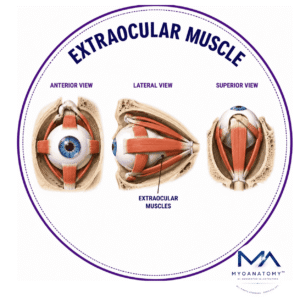
Extraocular Muscle

MYO CORE

Superior Oblique Muscle

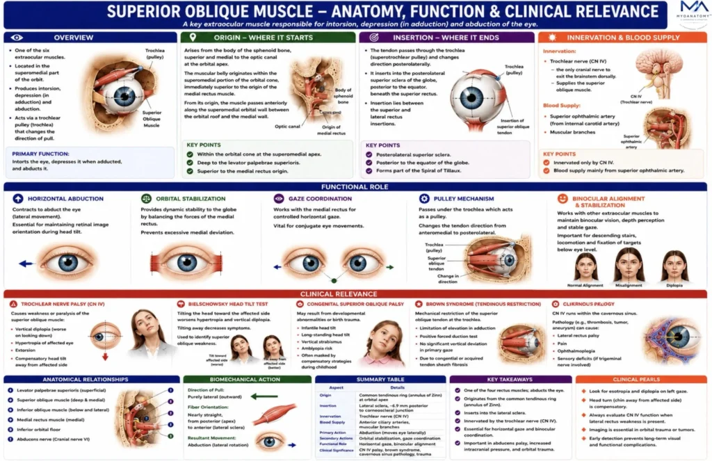
The superior oblique muscle is one of the six extraocular muscles responsible for controlling the orientation and rotation of the globe within the orbit. It belongs to the oblique group of extraocular muscles, characterized by their indirect course and their important role in ocular torsion and vertical gaze control

OVERVIEW

Unlike the rectus muscles, which act primarily in the cardinal planes, the superior oblique produces a complex three-dimensional rotational effect on the globe. This complexity results from the presence of the trochlea, a fibrocartilaginous pulley that redirects the direction of muscle contraction. As a consequence, the muscle generates a composite movement consisting of intorsion, depression, and abduction.

Innervated by the trochlear nerve (CN IV), the superior oblique plays a crucial role in torsional control of the eyeball, downward gaze, and stabilization of the visual field during head movement.

Dysfunction of the muscle produces characteristic vertical diplopia and compensatory head tilt, making it an important structure in both neurological and ophthalmological examination.

Functionally, the superior oblique plays a critical role in:

stabilization of the visual field during head tilt

downward gaze when the eye is adducted

torsional control of the eyeball

binocular coordination during locomotion and reading

The muscle receives motor innervation from the trochlear nerve (cranial nerve IV).

ANATOMY

Origin

The superior oblique arises from the:

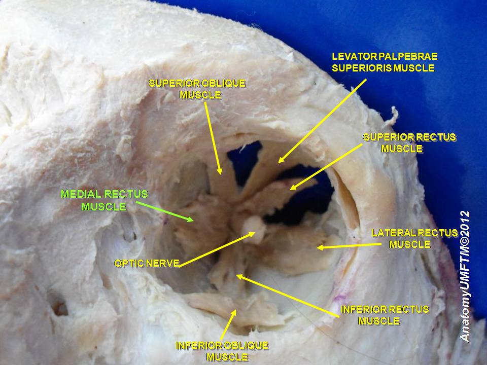
body of the sphenoid bone, superior and medial to the optic canal at the orbital apex.

The muscular belly originates within the superomedial portion of the orbital cone, immediately superior to the origin of the medial rectus muscle.

From this origin, the muscle passes anteriorly along the superior medial wall of the orbit, running between the:

orbital roof (frontal bone)

medial orbital wall (ethmoid bone)

The muscle belly gradually narrows into a tendinous portion before reaching the trochlea.

Trochlea (Pulley Apparatus)

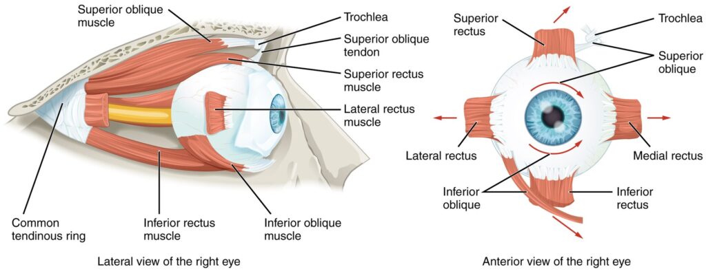
The tendon of the superior oblique passes through a fibrocartilaginous pulley known as the trochlea, located at the superomedial corner of the orbit on the frontal bone.

Exam Question

Evaluate the anatomical significance of the superior oblique origin from the sphenoid bone and explain how its superomedial orbital course influences the muscle’s mechanical advantage and subsequent actions on the globe

Insertion

After passing through the trochlea, the tendon travels:

posteriorly; laterally; superiorly

The tendon then inserts into the:

posterolateral superior sclera of the globe –  insertion is located posterior to the equator of the eyeball, beneath the superior rectus muscle.

Because the insertion lies posteror to the globe’s equator, contraction produces a rotational torque rather than simple linear movement.

Exam Question

Explain how the unique posterolateral scleral insertion of the superior oblique tendon, posterior to the equator of the globe, determines its rotational and depressor functions compared with the rectus muscles

Structural Characteristic

The trochlea is – a fibrocartilaginous ring anchored to the trochlear fossa of the frontal bone  lined by a synovial sheath that minimizes friction.

The tendon changes direction at the trochlea by approximately 45–55 degrees, redirecting its pull from anterior–posterior to posteromedial–lateral.

This pulley mechanism dramatically alters the vector of muscular force, allowing the muscle to generate rotational movements that would otherwise be impossible.

Exam Question

Analyze the biomechanical role of the trochlea as a fibrocartilaginous pulley and discuss how alteration of tendon direction at the trochlea modifies the vector of force transmitted to the globe.

Fascial & Pulley System

The superior oblique tendon is enclosed by a fascial sheath that blends with Tenon’s capsule (fascia bulbi).

Important connective structures include:

trochlear sheath-a synovial-like membrane surrounding the tendon at the trochlea, reducing friction during repeated eye movement.

intermuscular septa-connective tissue sheets connecting extraocular muscle sheaths and maintaining proper muscle alignment.

orbital pulley system-modern orbital biomechanics recognizes that extraocular muscles operate through fibromuscular pulley structures embedded within orbital connective tissue.

The superior oblique pulley system stabilizes the muscle path and ensures accurate transmission of force to the globe.

Disruption of these pulleys may lead to strabismus.

Exam Question

Discuss the contribution of the trochlear sheath, Tenon’s capsule, and orbital pulley system to superior oblique function, and evaluate their importance in maintaining efficient ocular movement and biomechanical precision.

Anatomical Relations

Superior –  orbital roof (frontal bone)

Inferior – superior rectus muscle

Medial – medial orbital wall ; Ethmoid bone

Posterior – optic nerve  Ophthalmic artery

Anterior – trochlea attached to frontal bone

The tendon passes beneath the superior rectus muscle before inserting onto the sclera.

Biomechanics of Superior Oblique Contraction -because the tendon passes through the trochlea, the line of pull forms an oblique vector relative to the globe. The muscle therefore produces three simultaneous actions.

Exam Question

Evaluate the anatomical relationships of the superior oblique muscle and tendon within the orbit and discuss how these relationships influence both surgical approaches and potential pathological involvement.

Innervation

Innervation by – trochlear nerve (cranial nerve IV)

Trochlear nucleus -located in the dorsal midbrain at the level of the inferior colliculus.

Important features:

motor neurons cross the midline before exiting the brainstem therefore each trochlear nerve supplies the contralateral superior oblique muscle

Course of the trochlear nerve

emerges dorsally from the midbrain

decussates within the brainstem

travels around the brainstem in the subarachnoid space

passes through the cavernous sinus

enters the orbit via the superior orbital fissure.

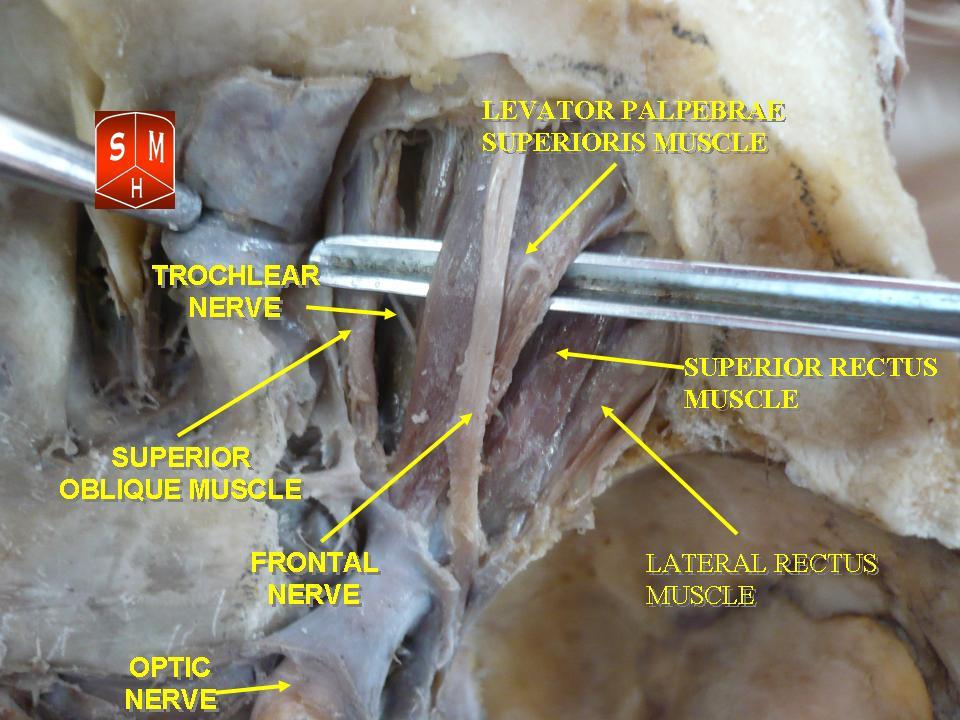
Within the orbit the nerve supplies the superior oblique on its superior surface.

Exam Question

Analyze the unique neuroanatomy of the trochlear nerve (CN IV), including its dorsal brainstem exit and complete decussation, and explain the clinical implications for superior oblique muscle dysfunction.

FUNCTIONAL ROLE

Intorsion Control

The superior oblique is the principal intortor of the eyeball, rotating the superior pole medially toward the nasal side. This torsional adjustment maintains retinal image orientation during head tilt and preserves spatial alignment of the visual field. 

Through its trochlear pulley mechanism, the muscle exerts highly efficient rotational control around the anteroposterior axis of the globe.

Exam Question

Explain why the superior oblique is considered the principal intortor of the eye and evaluate the importance of ocular intorsion in maintaining retinal image orientation during head movements.

Depressive Function

The superior oblique serves as an important depressor of the eye, particularly when the globe is adducted. In this position, the tendon assumes a more vertical vector of pull, maximizing downward movement. 

This biomechanical arrangement allows precise control of inferior gaze and complements the actions of the inferior rectus during complex ocular movements.

Exam Question

Analyze the biomechanical basis by which the superior oblique acts as a depressor of the adducted eye and compare this function with the depressor action of the inferior rectus muscle.

Vestibular Stabilization

The superior oblique contributes significantly to vestibulo-ocular reflex pathways that maintain visual fixation during head movement. 

During ipsilateral head tilt, coordinated activation of the superior oblique and contralateral extraocular muscles produces compensatory ocular torsion, ensuring that visual images remain stable on the retina despite changes in head position

Exam Question

Discuss the role of the superior oblique muscle in vestibulo-ocular reflex pathways and evaluate its contribution to visual fixation and retinal image stability during head motion.

Binocular Alignment

The muscle plays a critical role in maintaining binocular coordination, ocular balance, and dynamic gaze stabilization. Its actions are essential for downward visual tracking, reading, descending stairs, locomotion, and fixation of objects below eye level. 

By integrating torsional, vertical, and horizontal components of movement, the superior oblique supports accurate binocular vision and depth perception.

Exam Question

Evaluate how the superior oblique integrates torsional, vertical, and horizontal ocular movements to maintain binocular alignment, depth perception, and coordinated visual tracking.

CLINICAL RELEVANCE

Trochlear Palsy

Lesions of the trochlear nerve (CN IV) result in paralysis or weakness of the superior oblique muscle, producing impaired depression of the adducted eye, ocular extorsion, and vertical diplopia. 

Loss of normal torsional control disrupts binocular alignment, causing patients to adopt compensatory head postures to maintain single vision.

Exam Question

Analyze the pathophysiological mechanisms underlying trochlear nerve palsy and explain how superior oblique weakness produces vertical diplopia, extorsion, hypertropia, and compensatory head tilt.

Head Tilt Test

The Bielschowsky head tilt test is a fundamental clinical assessment used to identify superior oblique dysfunction. 

Tilting the head toward the affected side increases vertical misalignment and worsens diplopia, whereas tilting away reduces symptoms. This response reflects the inability of the weakened superior oblique to provide appropriate compensatory intorsion.

Exam Question

Evaluate the physiological basis of the Bielschowsky head tilt test and discuss its diagnostic value in identifying superior oblique dysfunction and trochlear nerve lesions.

Congenital Disfunction

Congenital superior oblique palsy commonly results from developmental abnormalities of the muscle, tendon, or trochlear nerve. 

Long-standing vertical strabismus, chronic compensatory head tilt, and impaired binocular vision are characteristic findings. Patients often develop adaptive mechanisms that may partially mask symptoms during early life.

Exam Question

Discuss the developmental mechanisms responsible for congenital superior oblique palsy and evaluate the adaptive compensatory strategies that may mask symptoms during childhood.

Brown Syndrome

Brown syndrome is a mechanical disorder caused by restriction of superior oblique tendon movement through the trochlea. Unlike neurogenic paralysis, ocular motility is limited by physical tethering of the tendon. 

The hallmark finding is inability to elevate the adducted eye, producing characteristic gaze limitation despite preserved neural innervation.

Exam Question

Analyze the anatomical and biomechanical basis of Brown syndrome and explain how restriction of superior oblique tendon movement through the trochlea produces characteristic limitations of ocular motility despite preserved neural innervation.

SUMMARY TABLE

Scroll to Top