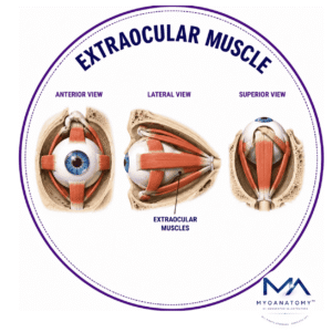
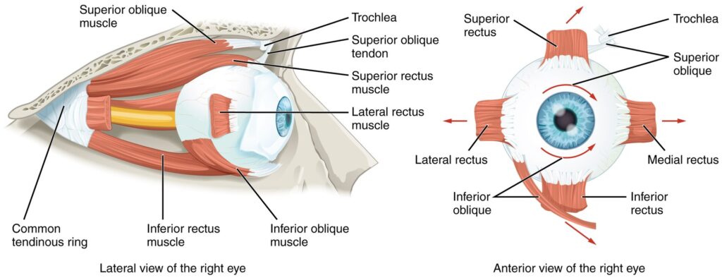
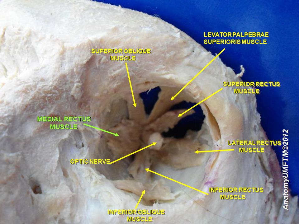
Extraocular Muscle
MYO CORE
Lateral Rectus Muscle
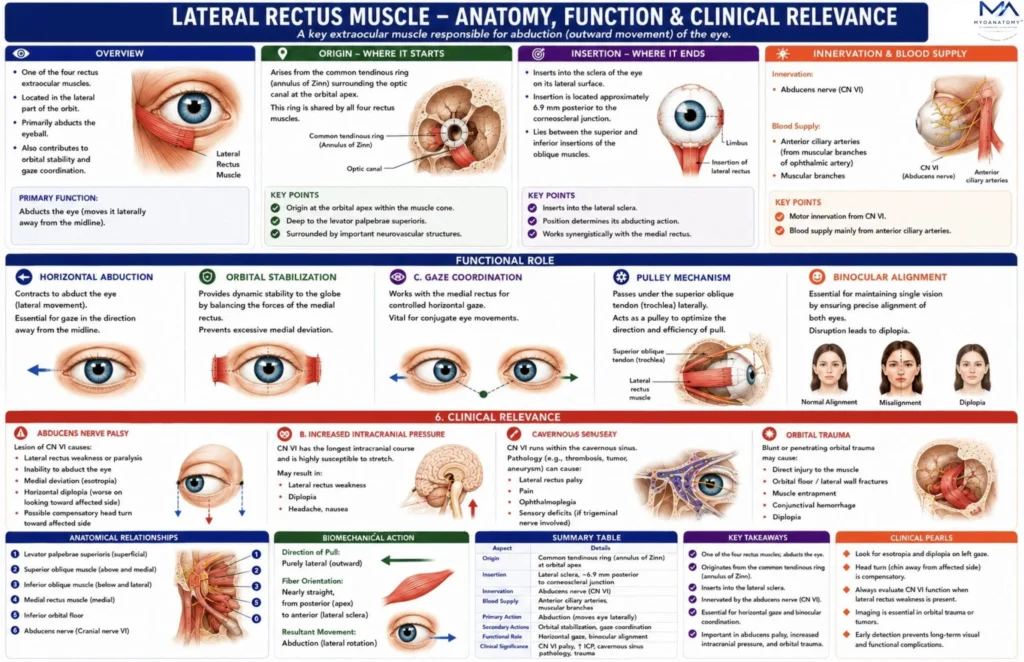
The lateral rectus muscle is one of the four rectus muscles of the orbit and serves as the primary abductor of the eyeball, moving the visual axis laterally away from the midline. It occupies the lateral aspect of the orbital cavity, extending from the common tendinous ring at the orbital apex to the lateral sclera of the globe
OVERVIEW
The lateral rectus muscle is a rectus extraocular muscle responsible for abduction of the eyeball. It originates from the common tendinous ring at the orbital apex and inserts into the lateral sclera of the globe. It is uniquely innervated by the abducens nerve (cranial nerve VI) and forms a key component of the horizontal gaze mechanism.
Through coordination with the contralateral medial rectus via brainstem gaze pathways, the lateral rectus enables conjugate horizontal eye movement, binocular vision, and visual tracking. Because of the vulnerability of the abducens nerve to intracranial pathology, dysfunction of the lateral rectus is a common and clinically important cause of diplopia and ocular misalignment.
The muscle is unique among extraocular muscles in that it is exclusively innervated by the abducens nerve (cranial nerve VI). Because the abducens nerve follows a long intracranial course across the clivus and cavernous sinus, the lateral rectus is particularly vulnerable to neurological pathology, making it one of the most clinically significant muscles in neuro-ophthalmology.


ANATOMY
Origin
The lateral rectus arises from the common tendinous ring (annulus tendineus communis or annulus of Zinn) located at the orbital apex.
The origin consists of two heads:
superior head – arises from the upper part of the tendinous ring
inferior head – arises from the lower part of the ring
Between these heads pass several critical structures entering the orbit:
optic nerve (CN II)
ophthalmic artery
oculomotor nerve divisions (CN III)
nasociliary nerve (branch of CN V1)
This anatomical arrangement forms part of the intraconal compartment of the orbit, through which many neurovascular structures travel.
From its origin, the muscle runs anteriorly along the lateral orbital wall, embedded within orbital fat.

Exam Question
Evaluate the anatomical origin of the lateral rectus muscle from the common tendinous ring (annulus of Zinn) and discuss the functional significance of its dual-headed origin, intraconal position, and relationship to the major neurovascular structures entering the orbit.
Insertion
The lateral rectus inserts into the:
lateral sclera anterior to the equator of the eyeball
The insertion is located approximately:
6.9–7 mm posterior to the corneoscleral junction
This insertion forms part of the Spiral of Tillaux, which describes the sequential insertion distances of the rectus muscles from the corneal limbus.
Order of insertion from anterior to posterior:
medial rectus (~5.5 mm)
inferior rectus (~6.5 mm)
lateral rectus (~6.9 mm)
superior rectus (~7.7 mm)
This spiral configuration has major importance in ocular alignment and strabismus surgery
Exam Question
Analyze the insertion of the lateral rectus muscle onto the sclera and explain the anatomical and clinical significance of its position within the Spiral of Tillaux, particularly in relation to ocular alignment, extraocular muscle biomechanics, and strabismus surgery.
Anatomical Relation
Fascial System and Orbital Pulley Mechanism
The lateral rectus is enclosed by the fascial sheath of the eyeball (Tenon’s capsule).
Several important fascial structures stabilize the muscle.
lateral check ligament -a fibrous extension from the lateral rectus sheath attaches to the lateral orbital wall near the zygomatic bone.
Exam Question
Evaluate the anatomical relationships of the lateral rectus muscle within the orbit and discuss the functional and clinical significance of its fascial attachments, orbital pulley system, and adjacent neurovascular structures.
Innervation
The lateral rectus receives motor innervation from:
abducens nerve (cranial nerve VI)
The nerve originates in the:
Abducens nucleus in the dorsal pons
It then follows a long intracranial course:
exits the brainstem at the pontomedullary junction
ascends along the clivus
passes through Dorello’s canal
traverses the cavernous sinus
enters the orbit via the superior orbital fissure
It then supplies the lateral rectus on its deep surface.
Exam Question
Analyze the complete anatomical course of the abducens nerve (CN VI) from its brainstem nucleus to the lateral rectus muscle and explain how lesions at different points along this pathway may produce characteristic ocular motor deficits.
FUNCTIONAL ROLE
Orbital Stabilization
The lateral rectus contributes to dynamic stabilization of the globe by balancing the adducting force of the medial rectus and maintaining proper ocular alignment within the orbit.
Through continuous interaction with the extraocular muscle system, it preserves binocular coordination, prevents excessive medial deviation, and supports stable visual fixation during gaze shifts.
Exam Question
Discuss the role of the lateral rectus muscle in maintaining ocular stability and evaluate its contribution to balanced extraocular muscle biomechanics and binocular alignment.
Pulley System
The lateral rectus operates through a fibroelastic orbital pulley that functions as a biomechanical guide, regulating the direction and efficiency of muscle pull.
This pulley system optimizes ocular rotation, stabilizes muscle position during gaze movements, and ensures precise horizontal control. Abnormal pulley positioning may alter ocular mechanics and contribute to complex forms of strabismus.
Exam Question
Evaluate the biomechanical significance of the lateral rectus pulley system and discuss its role in maintaining precise ocular alignment and horizontal gaze control
Horizontal Abduction
The lateral rectus is the principal abductor of the eye, producing lateral rotation of the globe away from the midline. Its fibers run almost parallel to the horizontal visual axis, allowing generation of a nearly pure horizontal movement with minimal vertical or torsional influence.
This unique orientation makes it the dominant muscle responsible for outward gaze.
Exam Question
Explain the anatomical and biomechanical factors that enable the lateral rectus muscle to function as the primary abductor of the eye.
Gaze Coordination
The lateral rectus is an essential component of the conjugate horizontal gaze network, acting in precise coordination with the contralateral medial rectus through the PPRF, abducens nucleus, and medial longitudinal fasciculus.
This integrated neural system synchronizes binocular eye movements, maintains single vision, and enables accurate tracking of moving targets.
Exam Question
Analyze the role of the lateral rectus muscle in conjugate horizontal gaze and discuss the neural mechanisms responsible for coordinated binocular eye movements
CLINICAL RELEVANCE
Abducens Nerve Palsy
The lateral rectus contributes to dynamic stabilization of the globe by balancing the adducting force of the medial rectus and maintaining proper ocular alignment within the orbit.
Through continuous interaction with the extraocular muscle system, it preserves binocular coordination, prevents excessive medial deviation, and supports stable visual fixation during gaze shifts.
Exam Question
Discuss the anatomical and functional consequences of abducens nerve palsy and explain how lateral rectus paralysis produces esotropia and horizontal diplopia.
Intracranial Pressure
The abducens nerve is particularly vulnerable to elevated intracranial pressure because of its long intracranial course and fixed anatomical attachments.
Increased pressure may stretch or compress the nerve, leading to unilateral or bilateral lateral rectus dysfunction. Consequently, abducens palsy is often considered an important localizing sign of intracranial hypertension.
Exam Question
Evaluate why the abducens nerve is especially susceptible to increased intracranial pressure and discuss the resulting effects on lateral rectus function and ocular motility.
Cavernous Sinus Pathology
The abducens nerve traverses the cavernous sinus in close association with the internal carotid artery and adjacent cranial nerves, making it vulnerable to vascular, inflammatory, infectious, and neoplastic lesions.
Pathology within this region may impair lateral rectus function and contribute to diplopia, ophthalmoplegia, and complex cranial neuropathies.
Exam Question
Analyze the anatomical relationship between the abducens nerve and the cavernous sinus and discuss the clinical manifestations of cavernous sinus lesions affecting lateral rectus function
Orbital Trauma
The lateral rectus may be affected by orbital fractures, inflammatory disorders, or soft-tissue injury involving the lateral orbit.
Mechanical restriction, muscle damage, or secondary fibrosis can impair abduction, disrupt binocular alignment, and produce diplopia during horizontal gaze.
Exam Question
Discuss the impact of orbital trauma and orbital disease on lateral rectus function and evaluate the mechanisms by which these conditions produce impaired abduction and diplopia.
SUMMARY TABLE