Suprahyoid Muscle

MYO CORE

Stylohyoid Muscle

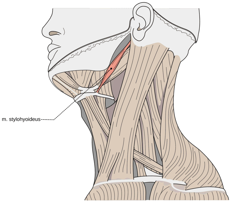
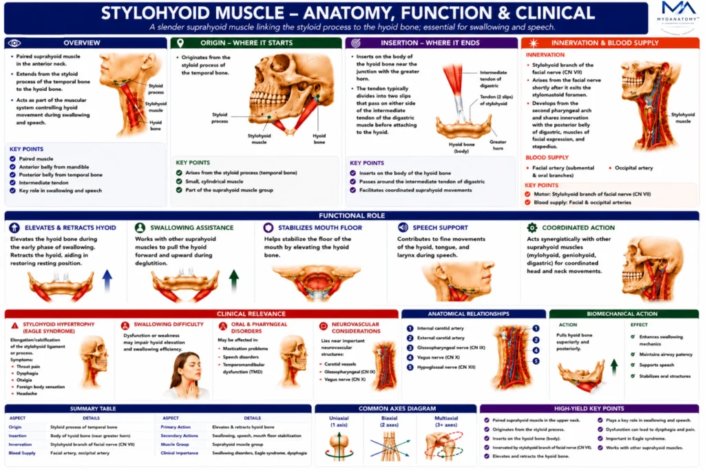
The stylohyoid muscle is a slender, paired suprahyoid muscle that extends from the styloid process of the temporal bone to the hyoid bone. It lies in the upper lateral region of the neck, forming part of the muscular system that controls hyoid movement during swallowing and speech.

OVERVIEW

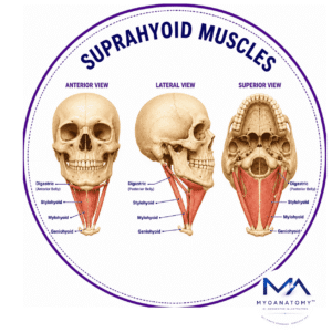
Functionally, the stylohyoid assists in elevating and retracting the hyoid bone, thereby contributing to the coordinated movements required for swallowing (deglutition) and stabilization of the floor of the mouth. The muscle acts together with the digastric, mylohyoid, and geniohyoid muscles as part of the suprahyoid muscular complex, which links the mandible, skull base, and hyoid apparatus.

A characteristic anatomical feature of the stylohyoid muscle is that its tendon splits near its insertion to allow passage of the intermediate tendon of the digastric muscle. This structural arrangement reflects the close functional relationship between these two muscles.

The stylohyoid muscle develops from the second pharyngeal (hyoid) arch and therefore receives motor innervation from the facial nerve (CN VII).

ANATOMY

Origin

Posterior surface of the styloid process of the temporal bone.

The styloid process is a slender projection of the temporal bone that serves as the attachment site for several muscles and ligaments associated with the hyoid apparatus and tongue

Exam Question

Evaluate how the attachment of the stylohyoid muscle to the styloid process contributes to hyoid biomechanics and upper cervical anatomy.

Insertion

The muscle fibers pass anteroinferiorly to insert on the:

body of the hyoid bone near the junction with the greater hor

Near its insertion, the tendon typically divides into two slips, which pass on either side of the intermediate tendon of the digastric muscle before attaching to the hyoid bone.

Exam Question

Discuss the insertion of the stylohyoid muscle and explain the anatomical and functional significance of its bifurcated tendon surrounding the intermediate tendon of the digastric muscle.

Innervation

The stylohyoid muscle is innervated by the:

stylohyoid branch of the facial nerve (CN VII)

This branch arises from the facial nerve shortly after it exits the stylomastoid foramen.

Because it develops from the second pharyngeal arch, the stylohyoid shares its innervation with other second-arch muscles such as:

posterior belly of digastric

muscles of facial expression

stapedius muscle.

Exam Question

Correlate the second pharyngeal arch origin of the stylohyoid muscle with its innervation pattern and associated muscular derivatives.

FUNCTIONAL ROLE

Hyoid Elevation & Retraction

The stylohyoid muscle elevates and retracts the hyoid bone during swallowing, contributing to coordinated movement of the hyoid–laryngeal complex. During the early phase of deglutition, elevation of the hyoid assists laryngeal ascent, airway protection, and efficient passage of the food bolus into the pharynx. 

As swallowing progresses, the stylohyoid helps retract the hyoid posteriorly, restoring the hyoid–laryngeal apparatus toward its resting position and preparing it for subsequent functional activity.

Exam Question

Evaluate the biomechanical significance of stylohyoid-mediated hyoid elevation and retraction, and discuss how these actions contribute to laryngeal movement, airway protection, and efficient swallowing physiology.

Speech Support

The stylohyoid muscle indirectly supports speech production by stabilizing and positioning the hyoid bone within the suprahyoid complex. 

By helping maintain optimal hyoid–laryngeal alignment, the stylohyoid contributes to precise control of vocal tract mechanics, facilitating clear speech articulation, normal resonance, and efficient coordination between respiratory, laryngeal, and oral structures during verbal communication.

Exam Question

Assess the contribution of the stylohyoid muscle to speech production and discuss how hyoid stabilization influences tongue mobility, laryngeal positioning, phonation, and articulation.

Swallowing Coordination

Acting synergistically with the posterior belly of the digastric muscle, the stylohyoid contributes to hyoid stabilization, pharyngeal widening, laryngeal elevation, and efficient bolus propulsion toward the esophagus. 

Together, these muscles form an essential component of the suprahyoid swallowing mechanism.

Exam Question

Evaluate the coordinated actions of the stylohyoid, digastric, mylohyoid, and geniohyoid muscles during swallowing, and explain how their integrated activity facilitates hyoid stabilization, laryngeal elevation, pharyngeal widening, and efficient bolus propulsion.

Stabilize Mouth Floor

The stylohyoid muscle contributes to stabilization of the floor of the mouth by maintaining the position of the hyoid bone and supporting the coordinated activity of the suprahyoid musculature. 

Through its attachment between the styloid process and hyoid bone, it helps establish a stable anatomical foundation upon which the tongue and floor-of-mouth muscles can function efficiently.

Exam Question

Analyze the contribution of the stylohyoid muscle to stabilization of the floor of the mouth and discuss its importance in tongue mobility, oral bolus control, and swallowing mechanics.

CLINICAL RELEVANCE

Stylohyoid Hypertrophy

Hypertrophy or abnormal enlargement of the stylohyoid muscle may alter normal hyoid biomechanics and contribute to discomfort during swallowing or neck movement. 

Although uncommon, excessive muscle bulk can interfere with coordinated suprahyoid function and may be associated with chronic muscular overuse or compensatory adaptations within the upper cervical region.

Exam Question

Evaluate the potential biomechanical consequences of stylohyoid hypertrophy and discuss its effects on hyoid movement and upper aerodigestive tract function.

Swallowing Difficulty

Dysfunction of the stylohyoid muscle may impair hyoid elevation and retraction during swallowing, reducing the efficiency of bolus transport through the pharynx. 

Patients may experience swallowing difficulty, delayed deglutition, or a sensation of impaired throat clearance, particularly when suprahyoid muscle coordination is compromised.

Exam Question

Analyze how stylohyoid dysfunction may contribute to swallowing difficulty and evaluate its role in normal deglutition biomechanics.

Oropharyngeal Disorders

Because the stylohyoid participates in coordinated movements of the hyoid, pharynx, and larynx, abnormalities affecting the muscle may contribute to oropharyngeal dysfunction.

Impaired muscle performance can disrupt normal swallowing mechanics, speech-related movements, and upper aerodigestive tract coordination, particularly in patients with neuromuscular or structural disorders.

Exam Question

Assess the contribution of the stylohyoid muscle to normal oropharyngeal function and discuss the clinical implications of its dysfunction.

Neurovascular Consideration

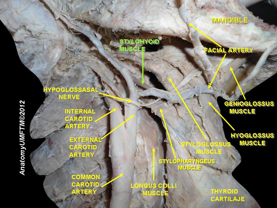
The stylohyoid muscle lies in close proximity to important neurovascular structures, including the facial nerve, external carotid artery, internal carotid artery, and internal jugular vein. 

Consequently, pathology or surgical intervention in this region requires careful anatomical awareness to avoid neurovascular injury and preserve normal cervical function.

Exam Question

Evaluate the neurovascular relationships of the stylohyoid muscle and discuss their significance in head and neck surgery and regional pathology.

SUMMARY TABLE

Scroll to Top