Extraocular Muscle
MYO CORE
Medial Rectus Muscle
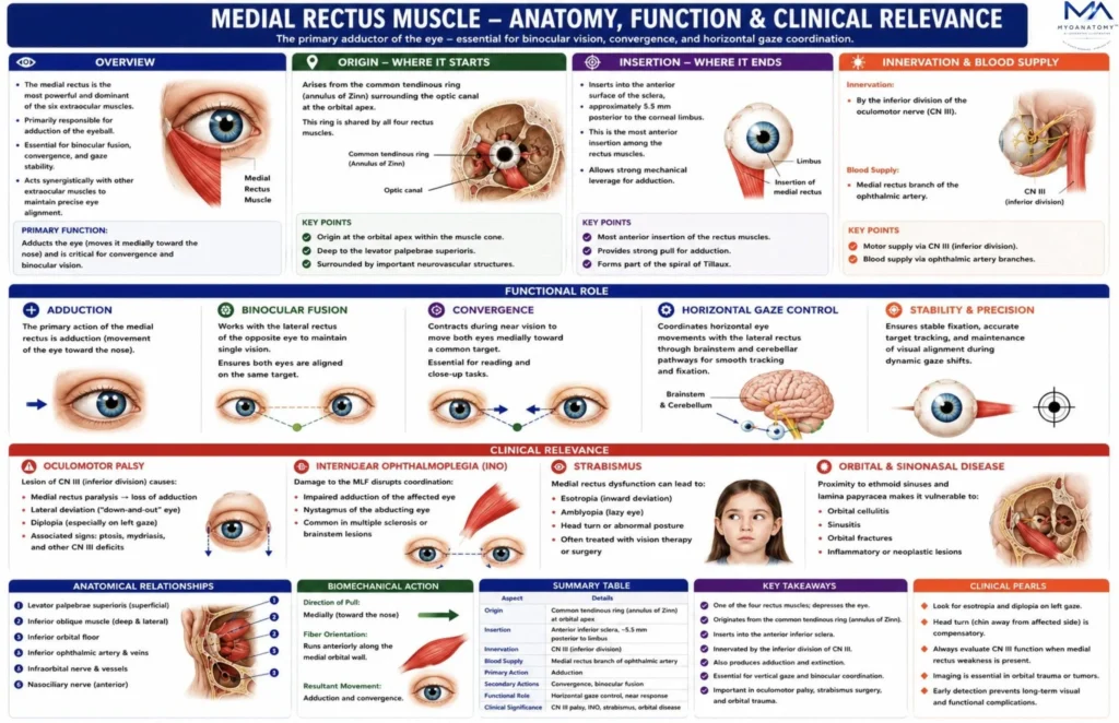
The medial rectus is the most powerful and functionally dominant of the six extraocular muscles. It is responsible for adduction of the eyeball, moving the visual axis toward the nasal midline. This movement is essential for binocular convergence, coordinated horizontal gaze, and precise alignment of the visual axes required for stereoscopic vision.
OVERVIEW
The medial rectus is the largest and most powerful extraocular muscle and is the primary adductor of the eyeball .
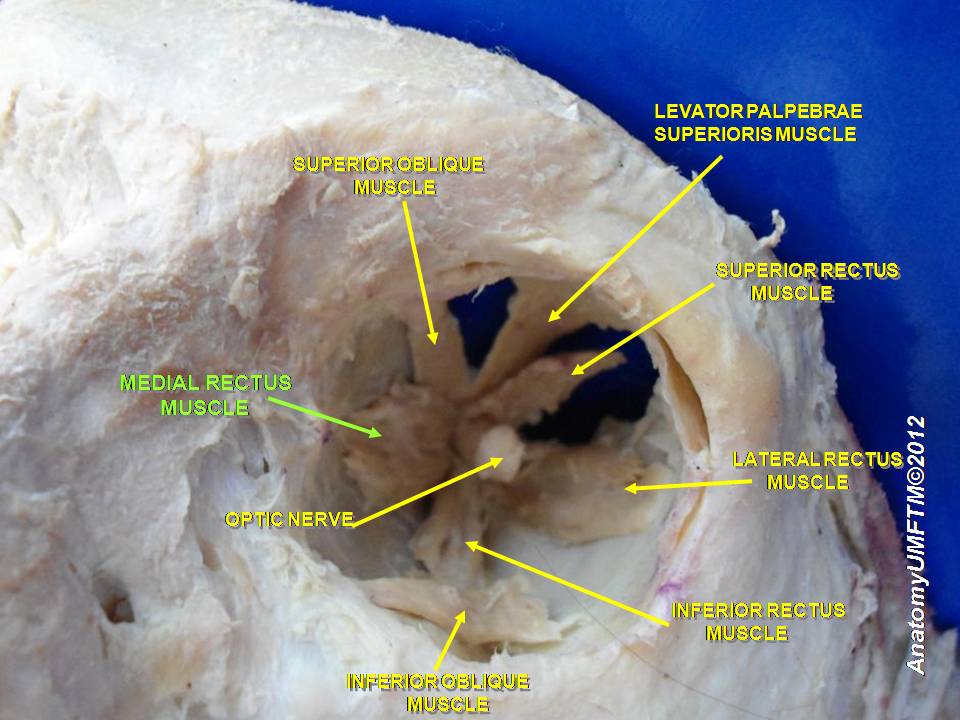
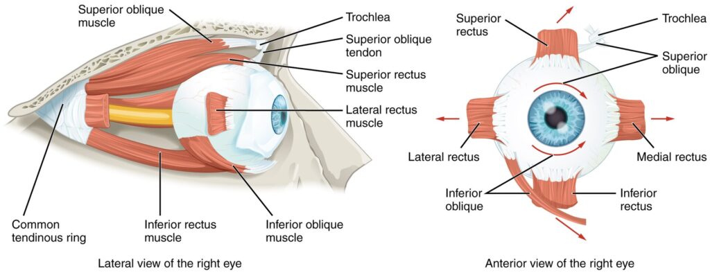
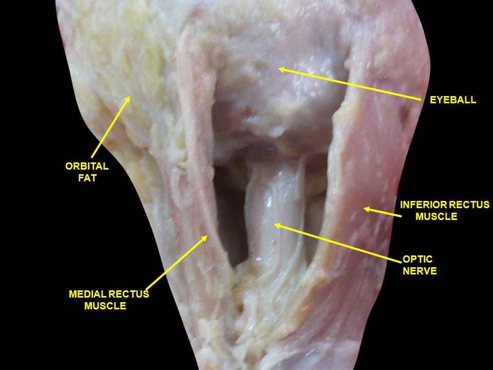
The muscle extends from the common tendinous ring (annulus of Zinn) at the orbital apex to the medial anterior sclera of the eyeball. It lies along the medial orbital wall, closely related to the thin lamina papyracea of the ethmoid bone. Because the medial wall is extremely thin, pathology arising from the ethmoid sinuses can easily affect structures adjacent to the medial rectus.
Among the extraocular muscles, the medial rectus has the largest physiological cross-sectional area, explaining its strong mechanical effect on the globe. The predominance of this muscle reflects the fundamental importance of convergence movements during near vision, which occur constantly during reading, writing, and interaction with nearby objects.
Functionally, the medial rectus forms part of the horizontal gaze system, which coordinates movements of both eyes so that they move together in a conjugate fashion. Its activity is tightly integrated with the contralateral lateral rectus through brainstem pathways, particularly the medial longitudinal fasciculus (MLF).
Clinically, the medial rectus is important in oculomotor nerve palsy, internuclear ophthalmoplegia, strabismus, and orbital disease, where even small disturbances in its function may lead to diplopia and impaired binocular vision

ANATOMY
Origin
The medial rectus arises from the:
common tendinous ring (annulus tendineus communis)
This ring surrounds:
optic canal; medial part of the superior orbital fissure
The annulus of Zinn provides the origin for the four rectus muscles:
superior rectus; inferior rectus
medial rectus; lateral rectus
From this origin the muscle travels anteriorly along the medial orbital wall, embedded within orbital fat.
Exam Question
Discuss the anatomical significance of the common tendinous ring as the origin of the medial rectus muscle.
Insertion
The medial rectus inserts into the:
medial sclera anterior to the equator of the eyeball
Its insertion lies approximately:
5.5 mm posterior to the corneal limbus
This is the most anterior insertion among the rectus muscles, providing strong mechanical leverage for adduction.
The insertion forms part of the spiral of Tillaux, which describes the progressive distances of rectus insertions frm the cornea.
Order of insertions from anterior to posterior:
Medial rectus (~5.5 mm)
Inferior rectus (~6.5 mm)
Lateral rectus (~6.9 mm)
Superior rectus (~7.7 mm)
This spiral arrangement is important in strabismus surgery.
Exam Question
Analyze the functional importance of the medial rectus insertion on the sclera and its relationship to the Spiral of Tillaux.
Anatomical Relation
Fascial System and Mechanical Support
like all extraocular muscles, the medial rectus is surrounded by Tenon’s capsule (fascia bulbi).
Important fascial structures include:
medial Check Ligament
a fibrous expansion of the medial rectus sheath attaches to the medial orbital wall.
Exam Question
Evaluate the key anatomical relationships of the medial rectus muscle and their clinical significance.
Innervation
The medial rectus receives motor innervation from:
inferior division of the oculomotor nerve (cranial nerve III)
The oculomotor nerve enters the orbit through the superior orbital fissure and divides into superior and inferior branches.
The inferior division supplies:
medial rectus
inferior rectus
inferior oblique
This nerve also carries parasympathetic fibers to the ciliary ganglion, which control pupil constriction and lens accommodation.
Exam Question
Describe the innervation of the medial rectus muscle and discuss the effects of oculomotor nerve lesions.
FUNCTIONAL ROLE
Adduction
The medial rectus is the primary adductor of the eye, producing medial rotation of the globe toward the midline. This action is fundamental for horizontal gaze, convergence, binocular alignment, and maintenance of single stereoscopic vision. Owing to its powerful mechanical leverage, it serves as the principal muscle responsible for precise near fixation and coordinated ocular movements.
Exam Question
Explain why the medial rectus is considered the principal adductor of the eye and discuss its importance in convergence, binocular alignment, and maintenance of single stereoscopic vision.
Binocular Vision
The medial rectus plays a central role in binocular vision by maintaining precise alignment of both visual axes on the same target. Through coordinated action with the contralateral lateral rectus, it enables single binocular vision, supports stereoscopic depth perception, and prevents diplopia during both distant and near fixation.
Exam Question
Discuss the role of the medial rectus muscle in binocular vision and explain how coordinated ocular alignment contributes to single vision, stereopsis, and depth perception
Near Response
The medial rectus serves as the principal effector of convergence within the near-response triad. Simultaneous contraction of both medial recti aligns the visual axes on a near object while accommodation and pupillary constriction occur concurrently.
This integrated neuro-ophthalmic mechanism is essential for sustained near fixation, reading, fine motor tasks, and detailed visual analysis.
Exam Question
Analyze the role of the medial rectus muscle in the near-response (accommodation) triad.
Conjugate Gaze
The medial rectus is a critical component of conjugate horizontal gaze, functioning synchronously with the contralateral lateral rectus through pathways involving the PPRF, abducens nucleus, and medial longitudinal fasciculus.
This highly coordinated neural network ensures smooth horizontal eye movements, stable fixation, accurate target tracking across the midline, and maintenance of binocular visual alignment during dynamic gaze shifts.
Exam Question
Evaluate the role of the medial rectus muscle in coordinated horizontal gaze movements.
CLINICAL RELEVANCE
Oculomotor Palsy
Lesions of the oculomotor nerve (CN III) produce paralysis of the medial rectus, resulting in loss of adduction and disruption of normal binocular alignment.
The unopposed actions of the lateral rectus and superior oblique muscles displace the globe laterally and inferiorly, producing the characteristic “down-and-out” position accompanied by horizontal diplopia and impaired conjugate gaze.
Exam Question
Discuss the anatomical and functional consequences of medial rectus paralysis in oculomotor nerve palsy.
Internuclear Ophtalmoplegia
Internuclear ophthalmoplegia (INO) results from damage to the medial longitudinal fasciculus (MLF), the critical brainstem pathway coordinating activity between the medial rectus and contralateral lateral rectus.
Patients demonstrate impaired adduction during horizontal gaze with abducting nystagmus of the opposite eye, while convergence remains preserved. This dissociation represents a classic neuro-ophthalmological finding commonly associated with multiple sclerosis and brainstem infarction.
Exam Question
Explain the pathophysiological basis of impaired medial rectus function in internuclear ophthalmoplegia.
Strabismic Disorder
Imbalance between the medial rectus and lateral rectus disrupts ocular alignment and may result in horizontal strabismus. Excessive medial rectus activity or lateral rectus weakness produces esotropia, whereas medial rectus weakness or lateral rectus overaction leads to exotropia.
These disorders impair binocular fusion, reduce stereopsis, and may generate diplopia or suppression of visual input from one eye.
Exam Question
Analyze the role of the medial rectus muscle in the development of horizontal strabismus.
Orbital Pathology
Because the medial rectus lies immediately adjacent to the ethmoidal air cells and thin lamina papyracea, it is particularly vulnerable to orbital and sinonasal disease.
Orbital cellulitis, ethmoidal sinus infection, inflammatory processes, and medial orbital wall fractures may directly affect muscle function, producing painful ocular movement, restricted adduction, diplopia, and disturbances of binocular vision.
Exam Question
Evaluate the clinical significance of medial rectus involvement in orbital and sinonasal disease.
SUMMARY TABLE