Lacrimal Bone

The lacrimal bones are the smallest and most delicate paired bones of the viscerocranium, located in the anteromedial wall of the orbit. They lie posterior to the frontal process of the maxilla and anterior to the ethmoid bone, forming part of the medial orbital wall and contributing to the lacrimal apparatus responsible for tear drainage

CORE

Overview

Despite their small size, the lacrimal bones play an essential role in the lacrimal drainage system, which conducts tears from the lacrimal sac into the nasal cavity. Structurally, they participate in the formation of the lacrimal fossa, which accommodates the lacrimal sac, a component of the tear drainage pathway.

Through their articulations with surrounding bones of the orbit and nasal cavity, the lacrimal bones contribute to the structural stability of the medial orbital wall while simultaneously linking the orbit to the nasal cavity through the nasolacrimal duct system.

ANATOMY

Surfaces

Each lacrimal bone presents 2  principal surfaces, corresponding to its relationships with the orbit and the nasal cavity.

Orbital Surface –  forms part of the medial wall of the orbit. This surface contains the posterior lacrimal crest, which serves as an attachment for the lacrimal fascia. Anterior to this crest lies the lacrimal groove, which contributes to the lacrimal fossa together with the frontal process of the maxilla. This fossa accommodates the lacrimal sac, which collects tears from the lacrimal canaliculi before transmitting them into the nasolacrimal duct. The orbital surface therefore plays an important role in the structural organization of the lacrimal drainage system.

Nasal Surface–  contributes to the lateral wall of the nasal cavity, forming part of the boundary between the orbit and nasal passages. Through its relationship with the nasolacrimal duct, the lacrimal bone participates in the anatomical pathway that allows tears to drain from the eye into the inferior nasal meatus.

Exam Question

Analyze how differences between the orbital and nasal surfaces of the lacrimal bone reflect their roles in orbital support and lacrimal drainage, and how disruption affects tear flow and orbital–nasal separation.

Borders

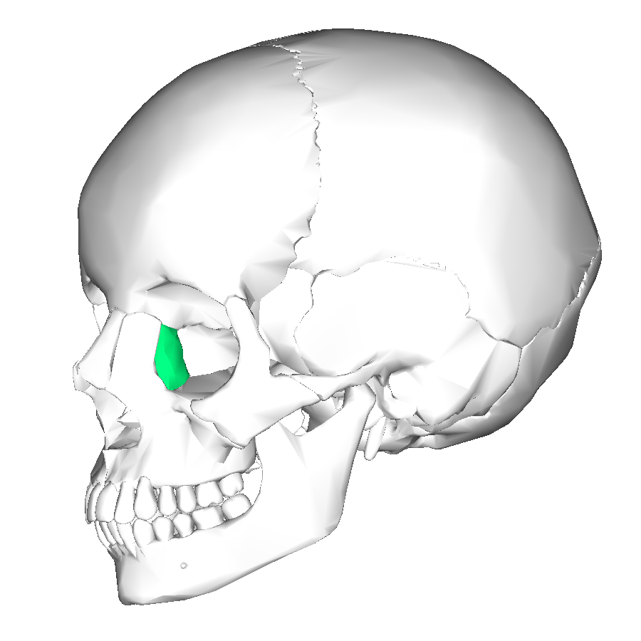
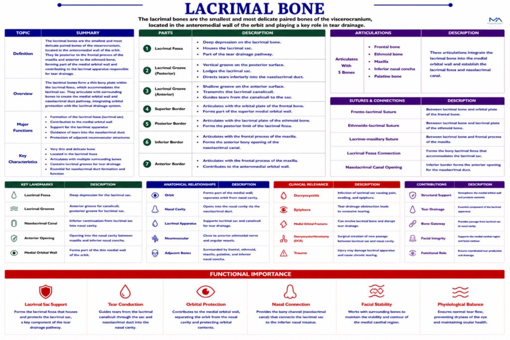
Each lacrimal bone presents 4  borders, which articulate with surrounding bones and integrate the lacrimal bone into the orbital and nasal skeletal framework.

Superior Border –  articulates with the orbital plate of the frontal bone, contributing to the medial orbital wall.

Inferior Border –  articulates with the maxilla, helping form the nasolacrimal canal, which transmits the nasolacrimal duct.

Posterior Border– articulates with the ethmoid bone, specifically with the lamina papyracea, contributing to the medial orbital wall.

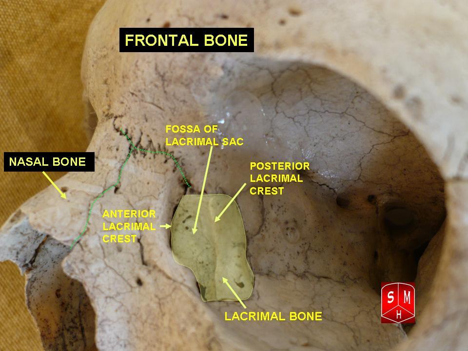
Anterior Border-  articulates with the frontal process of the maxilla, completing the lacrimal fossa.

Exam Question

Evaluate how the borders of the lacrimal bone contribute to medial orbital wall stability and the nasolacrimal system, and the consequences of their disruption in facial trauma.

Articulations

Each lacrimal bone articulates with 4 bones:

frontal bone

ethmoid bone

maxilla

inferior nasal concha

Through these articulations, the lacrimal bone forms part of both the orbital cavity and the nasal cavity, linking the ocular and respiratory systems.

Exam Question

Discuss how lacrimal bone articulations integrate the orbit and nasal cavity, and their role in coordinating tear drainage and nasal function.

Landmarks

Important anatomical landmarks of the lacrimal bone include:

Posterior lacrimal crest – attachment for lacrimal fascia

Lacrimal groove – contributes to formation of lacrimal fossa

Lacrimal fossa – accommodates the lacrimal sac

Nasolacrimal canal (with maxilla)  – passage for the nasolacrimal duct.

These structures form part of the lacrimal drainage apparatus, which transports tears from the eye to the nasal cavity

Exam Question

Assess the clinical significance of lacrimal bone landmarks in tear drainage, imaging interpretation, and surgical procedures such as dacryocystorhinostomy.

FUNCTIONAL IMPORTANCE

Lacrimal Drainage


The lacrimal bone forms the bony lacrimal fossa, which houses the lacrimal sac – an essential component of the tear drainage pathway. 

It provides the rigid scaffold guiding tears from the canaliculi into the nasolacrimal duct, ensuring unidirectional tear flow from the ocular surface to the inferior nasal meatus.

Orbital Wall Stability

As part of the medial orbital wall, the lacrimal bone contributes to the containment and protection of orbital contents, including the globe and extraocular muscles. 

Despite its thinness, it acts as a structural interface between the orbit and adjacent ethmoidal air cells.

Nasolacrimal Interface

The lacrimal bone forms a critical junction between the orbit and nasal cavity, coordinating ocular surface physiology with nasal airflow dynamics. 

This anatomical continuity explains the functional link between tear production and nasal mucosal hydration.

Nasal Framework

Through its articulation with the maxilla and ethmoid, the lacrimal bone contributes to the stability of the anterior facial skeleton, particularly supporting structures involved in nasal contour and medial canthal anatomy.

CLINICAL RELEVANCE

Orbital Fractures

The lacrimal bone, due to its extreme thinness, is highly susceptible to involvement in medial orbital (blow-out) fractures. Disruption of this region compromises the integrity of the medial orbital wall, allowing communication with adjacent ethmoidal air cells. 

This may result in orbital emphysema, diplopia, and impairment of lacrimal drainage, reflecting the bone’s role as a structural boundary between orbital and sinonasal compartments.

Drainage Obstraction

The lacrimal bone forms the lacrimal fossa, essential for alignment of the lacrimal sac within the tear drainage pathway. Trauma, inflammation, or structural displacement can obstruct this pathway, leading to epiphora (excessive tearing). 

This condition arises from failure of tear outflow into the nasolacrimal duct, highlighting the importance of precise anatomical positioning for maintaining functional patency

Lacrimal Infection

Obstruction of the nasolacrimal system predisposes to dacryocystitis, an infection of the lacrimal sac. Tear stasis promotes bacterial growth, resulting in painful swelling at the medial canthus, erythema, and purulent discharge. 

The lacrimal bone defines the anatomical site of this infection, emphasizing its role in localizing and facilitating pathological processes.

Surgical Access

The lacrimal bone is a key landmark in dacryocystorhinostomy (DCR), a procedure that creates a direct connection between the lacrimal sac and nasal cavity. Partial removal of the lacrimal bone allows bypass of an obstructed nasolacrimal duct. 

Accurate anatomical knowledge is critical to avoid damage to the orbit and surrounding structures, underscoring its importance in ophthalmic and ENT surgery

SUMMARY TABLE

Scroll to Top