Maxillae Bone
The maxillae are paired bones forming the central structural framework of the midface and the upper jaw. They are among the most functionally significant bones of the viscerocranium because they contribute simultaneously to the orbit, nasal cavity, oral cavity, and facial skeleton.
CORE


Overview
Each maxilla participates in the formation of the floor of the orbit, the lateral walls and floor of the nasal cavity, and the anterior portion of the hard palate, thereby linking the respiratory, digestive, and visual systems within a single structural complex. The maxillae also support the upper dental arch, which plays a major role in mastication and phonation.
Because of these multiple anatomical contributions, the maxilla represents a central structural hub of the facial skeleton, articulating with numerous cranial and facial bones and serving as an attachment site for muscles involved in facial expression and mastication.
ANATOMY
Processes
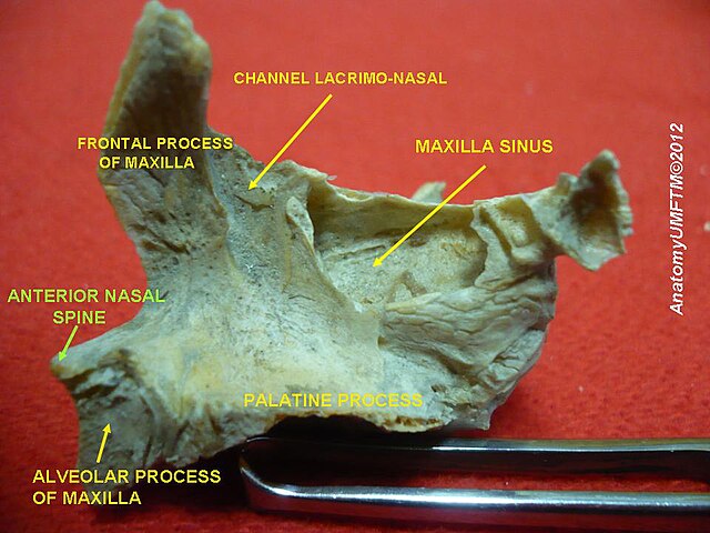
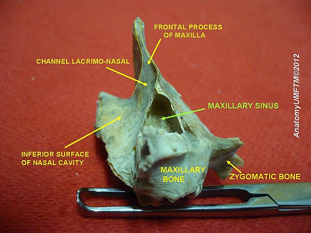
Each maxilla consists of a body and 4 processes:
Frontal Process – extends superiorly to articulate with the frontal bone and contributes to the medial wall of the orbit. It also forms part of the lateral boundary of the nasal cavity.
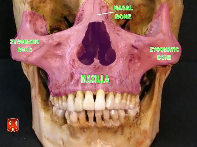
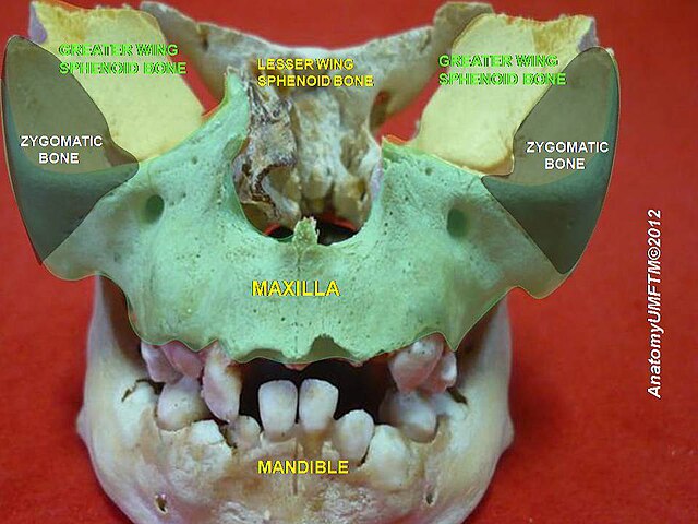
Zygomatic Process – extends laterally and articulates with the zygomatic bone, contributing to the lateral facial skeleton and orbital margin.
Palatine Process – of the two maxillae meet in the midline to form the anterior three-quarters of the hard palate, which separates the oral cavity from the nasal cavity. This structure is essential for speech and swallowing, allowing simultaneous breathing and eating.
Alveolar Process -contains the alveoli, which are sockets that house the roots of the upper teeth. This part of the bone adapts and remodels in response to tooth eruption and loss. These components allow the maxilla to contribute to several anatomical regions of the face.
Exam Question
Discuss how the frontal, zygomatic, palatine, and alveolar processes of the maxilla collectively organize the orbit, nasal cavity, oral cavity, and dentoalveolar apparatus, and explain how this arrangement permits simultaneous structural support, compartment separation, and transmission of occlusal forces through the midface.
Surfaces
The body presents 4 surfaces:
Anterior (facial) surface– forms the front of the upper jaw and midface.
Orbital surface- forms the major portion of the floor of the orbit. Running through this surface is the infraorbital groove, which continues anteriorly as the infraorbital canal and opens at the infraorbital foramen. This canal transmits the infraorbital nerve, a branch of the maxillary nerve (CN V₂).
Nasal Surface- forms part of the lateral wall of the nasal cavity. It contains the maxillary hiatus, a large opening that connects the maxillary sinus to the nasal cavity. In the articulated skull, this opening is partially closed by adjacent bones such as the palatine bone and inferior nasal concha.
Infratemporal Surface– contributes to the infratemporal fossa, an anatomical space containing important muscles of mastication and neurovascular structures. It contains small openings called alveolar foramina, which transmit the posterior superior alveolar nerves and vessels supplying the upper teeth.
Exam Question
Evaluate the functional anatomy of the maxillary surfaces by explaining how the facial, orbital, nasal, and infratemporal surfaces together integrate facial contour, orbital support, sinonasal drainage, and posterior alveolar neurovascular relations within a single skeletal unit.
Borders
The maxilla forms important borders of the facial skeleton.
Inferior border – contributes to the upper dental arch
Medial border – participates in the nasal cavity
Superior border – contributes to the orbital floor
Exam Question
Analyze the borders of the maxilla as structural transition zones between the dental arch, nasal aperture, and orbital floor, and explain how their arrangement maintains midfacial stability while linking mastication, respiration, and orbital integrity.
Articulations
The maxilla articulates with 9 bones, reflecting its central position within the facial skeleton.
These include:
Frontal bone ; Nasal bones
Zygomatic bones; Lacrimal bones
Ethmoid bone; Palatine bones
Inferior nasal concha; Vomer
Opposite maxilla
Through these articulations, the maxilla integrates the orbit, nasal cavity, and oral cavity into a unified anatomical framework.
Exam Question
The maxilla articulates with numerous facial and cranial bones. Explain how these articulations establish the maxilla as the central architectural hub of the viscerocranium, with particular emphasis on force redistribution, orbital–nasal–oral integration, and maintenance of midfacial projection.
Landmarks
Important anatomical landmarks include:
Infraorbital foramen – passage for infraorbital nerve and vessels
Maxillary sinus – largest paranasal sinus
Alveolar process – supports upper teeth
Anterior nasal spine –midline projection contributing to the inferior margin of the nasal aperture
Infraorbital canal – transmits the infraorbital nerve and vessels, supplying the lower eyelid, upper lip, and cheek
Canine fossae- depression above the canine tooth root
Exam Question
Discuss the major landmarks of the maxilla as clinically significant expressions of its internal organization, showing how the infraorbital canal and foramen, maxillary sinus, alveolar process, anterior nasal spine, and canine fossa reflect the convergence of neurovascular transmission, sinus physiology, dentition, and surgical access within the midface.
FUNCTIONAL IMPORTANCE
Structural Integration
The maxilla functions as the central load-bearing unit of the midface, integrating the oral, nasal, and orbital compartments.
Through its articulations, it redistributes masticatory forces across craniofacial buttress systems, maintaining structural stability and resistance to deformation
Occlusal Dynamics
The alveolar process anchors the maxillary dentition, enabling precise occlusal alignment with the mandible.
Repetitive mechanical loading during mastication drives continuous alveolar bone remodeling via mechanotransduction, ensuring functional adaptation and long-term skeletal integrity.
Palatal Partioning
The palatine processes form the anterior hard palate, creating a rigid separation between the oral and nasal cavities.
This enables coordinated respiration and feeding while providing a stable platform for tongue-driven articulation during swallowing and speech.
Air-Orbital Interface
The maxilla simultaneously forms the floor of the orbit and the lateral wall of the nasal cavity, acting as a structural interface between visual and respiratory systems.
It supports the globe, maintains orbital volume, and regulates airflow dynamics, ensuring functional compartmentalization and physiological efficiency.
CLINICAL RELEVANCE
Le Fort Fractures
Le Fort fractures represent progressive disruption of the midfacial buttress system (nasomaxillary, zygomaticomaxillary, pterygomaxillary), which normally transmits masticatory forces from the maxilla to the cranial base.
Loss of this structural continuity results in craniofacial instability, with combined impairment of occlusion, orbital support, and airway patency, reflecting failure of the integrated midfacial load-bearing framework.
Infraoribital Injury
The infraorbital nerve (CN V₂) courses within the infraorbital canal beneath the orbital floor, making it highly vulnerable in maxillary and orbital fractures (especially blowout injuries).
Compression or disruption produces hypoesthesia of the midface, with deficits in the upper lip, cheek, and lower eyelid, illustrating the sensitivity of confined neurovascular pathways to even minimal structural displacement or edema.
Sinus Pathology
The maxillary sinus maintains intimate anatomical relationships with the roots of the posterior maxillary teeth and drains into the middle nasal meatus.
Obstruction of this drainage pathway impairs mucociliary clearance, leading to secretion stasis and secondary infection, while odontogenic sources may directly extend into the sinus, demonstrating the functional integration of dental, nasal, and sinus systems.
Palatal Defects
Failure of fusion of the palatine processes during embryogenesis results in cleft palate, creating a persistent communication between oral and nasal cavities.
This disrupts functional compartmentalization, leading to impaired feeding, speech articulation, and airway control, and reflects the maxilla’s essential role in coordinated craniofacial development and oropharyngeal function.
SUMMARY TABLE
