Zygomatic Bone
The zygomatic bone is a paired bone of the viscerocranium forming the prominence of the cheek and contributing significantly to the lateral wall and floor of the orbit, the zygomatic arch, and the anterolateral facial skeleton. Because of its strategic position between the maxilla, frontal bone, temporal bone, and sphenoid, the zygomatic bone functions as a major structural pillar of the midface, transmitting mechanical forces between the facial skeleton and the cranial base.
CORE


Overview
Structurally, the zygomatic bone contributes to the orbital framework, the zygomatic arch, and the midfacial contour, thereby playing a central role in both craniofacial biomechanics and facial morphology.
It also transmits sensory branches of the maxillary division of the trigeminal nerve (CN V₂) and provides attachment sites for muscles involved in mastication and facial expression.
ANATOMY
Processes
The zygomatic bone presents 3 major processes that connect it with surrounding bones and stabilize the midface.
Frontal Process – extends superiorly and articulates with the frontal bone, forming part of the lateral orbital margin and contributing to the structural reinforcement of the superolateral orbit.
Temporal Process – projects posteriorly and articulates with the zygomatic process of the temporal bone, forming the zygomatic arch. This arch acts as a biomechanical bridge between the facial skeleton and cranial base.
Maxillary Process – extends medially and articulates with the maxilla, contributing to the inferolateral margin of the orbit and the midfacial framework.
Exam Question
Explain how the frontal, temporal, and maxillary processes of the zygomatic bone collectively establish a load-bearing framework of the midface, and analyze their role in redistributing masticatory forces to the cranial base.
Surfaces
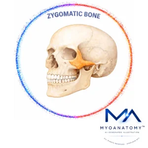
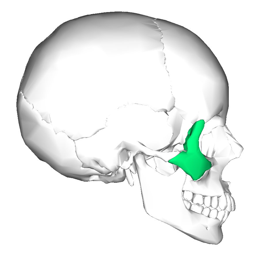
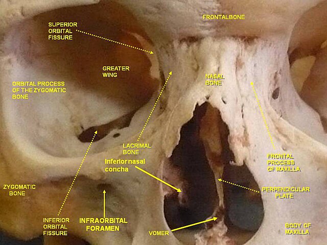
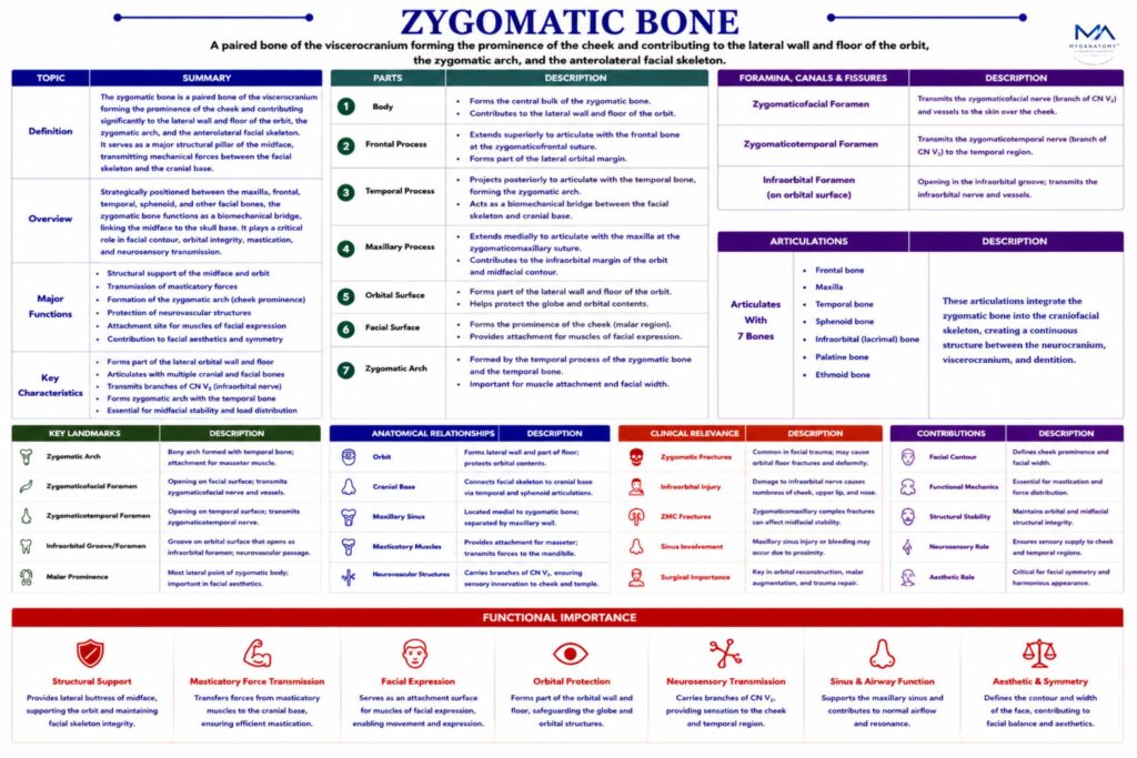
The zygomatic bone presents 3 principal surfaces, each contributing to different anatomical regions.
Facial Surface – forms the prominence of the cheek and provides attachment for muscles of facial expression. A notable landmark on this surface is the zygomaticofacial foramen, which transmits the zygomaticofacial nerve and vessels, supplying the skin of the cheek.
Temporal Surface – faces posteriorly and contributes to the temporal fossa and infratemporal fossa, regions containing important muscles of mastication and neurovascular structures. This surface contains the zygomaticotemporal foramen, transmitting the zygomaticotemporal nerve supplying the temporal region.
Orbital Surface – contributes to the lateral wall and floor of the orbit, helping form a strong lateral boundary that protects the globe and orbital contents from lateral mechanical forces. This surface contains the zygomatico-orbital foramen, which transmits branches of the zygomatic nerve within the orbital cavity.
Exam Question
Critically evaluate how the facial, temporal, and orbital surfaces of the zygomatic bone integrate muscular attachment, neurovascular transmission, and orbital protection, and explain how disruption of each surface alters regional function.
Borders
The zygomatic bone presents several borders that articulate with adjacent bones and form important structural landmarks.
Orbital border – contributes to the lateral margin of the orbit.
Maxillary border – articulates with the maxilla and forms part of the orbital rim
Temporal border – participates in the formation of the zygomatic arch
Frontal border – articulates with the frontal bone at the zygomaticofrontal suture
These borders integrate the zygomatic bone into the midfacial skeletal framework
Exam Question
Discuss how the borders of the zygomatic bone act as biomechanical junctions between cranial and facial skeleton, and analyze their role in maintaining structural continuity and resisting midfacial deformation under load.
Surfaces
Each zygomatic bone articulates with 4 bones:
Frontal bone
Maxilla
Temporal bone
Sphenoid bone
Through these articulations, the zygomatic bone connects the facial skeleton with the cranial base, stabilizing the orbital region and lateral midface.
Exam Question
Analyze how the articulations of the zygomatic bone with the frontal, maxillary, temporal, and sphenoid bones form an integrated craniofacial unit, and explain their importance in stabilizing the orbit and transmitting forces across the skull.
Landmarks
Important anatomical landmarks of the zygomatic bone include:
Zygomaticofacial foramen – passage for sensory nerves to the cheek
Zygomaticotemporal foramen – passage for nerves to the temporal region
Zygomatic arch – formed with the temporal bone
Orbital margin – structural boundary of the orbit
These landmarks are clinically important in orbital surgery and maxillofacial anatomy.
Exam Question
Evaluate the clinical significance of key zygomatic bone landmarks in relation to sensory nerve pathways, surgical access points, and the preservation of orbital and midfacial integrity.
FUNCTIONAL IMPORTANCE
Loud Bearing
The zygomatic bone functions as a primary lateral buttress of the midface, forming the zygomaticomaxillary and zygomaticotemporal load-bearing columns. It transmits masticatory forces from the maxilla toward the cranial base, redistributing stress across the lateral facial skeleton.
This structural role ensures resistance to lateral compression and shear forces, preserving orbital integrity and preventing midfacial collapse during high mechanical loading.
Mastication
Through formation of the zygomatic arch (with the temporal bone), the zygomatic bone provides a critical attachment site for the masseter muscle, a principal elevator of the mandible.
This converts muscular contraction into efficient bite force generation, acting as a biomechanical bridge between the facial skeleton and mandibular movement, thereby optimizing chewing efficiency and occlusal stability.
Orbit Stabilization
The zygomatic bone forms a substantial portion of the lateral orbital wall and inferolateral rim, contributing to the rigid osseous framework that protects the globe.
Its geometry absorbs and dissipates external forces away from the orbit, maintaining orbital volume, globe position, and neurovascular protection, which is essential for preserving visual function under mechanical stress.
Midfacial Projection
The zygomatic bone defines the anterior projection and contour of the midface, influencing facial symmetry and soft tissue distribution.
It also transmits sensory branches of CN V₂ (zygomaticofacial and zygomaticotemporal nerves), integrating skeletal structure with cutaneous innervation. This reflects its dual role in aesthetic architecture and neurofunctional mapping of the face.
CLINICAL RELEVANCE
Zygomaticomaxillary Disruption
ZMC (tripod) fractures separate the zygoma at the zygomaticofrontal suture, zygomaticomaxillary junction, and zygomatic arch, detaching it from its principal articulatory pillars.
This collapses the lateral midfacial buttress system, disrupting transmission of occlusal forces to the cranial base and producing mechanical decoupling of the midface.
Clinically, this results in malar depression, facial asymmetry, and orbital rim instability, reflecting failure of the zygoma as a key biomechanical stabilizer.
Infraoribital Injury
The infraorbital nerve (CN V₂) courses within a confined bony canal beneath the orbital floor, making it highly susceptible in ZMC fractures.
Fracture-related edema or displacement produces compressive neuropathy, causing hypoesthesia of the cheek, upper lip, and lower eyelid.
This illustrates how non-compliant osseous canals amplify minor structural changes into significant neural dysfunction.
Orbital Failure
The zygoma forms the lateral orbital wall and inferolateral rim, maintaining orbital volume and geometric stability.
Fracture increases orbital volume, leading to:
Enophthalmos
Diplopia
Herniation of orbital contents
These changes reflect loss of three-dimensional orbital integrity and load distribution, compromising both mechanical support and visual alignment
Mastication Disfunction
The zygomatic arch acts as a force-transducing lever, transmitting masseter and temporalis forces to the mandible.
Fracture disrupts this pathway and may obstruct the coronoid process, resulting in:
Reduced bite force
Painful mastication
Trismus
This demonstrates the zygoma’s role as a dynamic interface between craniofacial structure and mandibular mechanics.
SUMMARY TABLE
