Palatine Bone

The palatine bones are paired -shaped bones located in the posterior region of the viscerocranium, forming an essential component of the hard palate, nasal cavity, orbit, and pterygopalatine fossa. Although relatively small, these bones play a significant role in the structural integration of the midface and cranial base, linking the oral cavity, nasal cavity, orbit, and deep facial spaces

CORE

Overview

Each palatine bone lies posterior to the maxilla, inferior to the sphenoid bone, and medial to the pterygopalatine fossa, occupying a strategic anatomical position where several craniofacial compartments intersect. Through its articulations with multiple cranial and facial bones, the palatine bone contributes to the architecture of the posterior hard palate, the lateral wall of the nasal cavity, and a small portion of the orbital floor.

Palatine bones help maintain the structural separation between the oral and nasal cavities, support respiratory airflow pathways within the nasal cavity, and form part of the framework of the pterygopalatine region,

ANATOMY

Parts

Each palatine bone consists of 2  primary plates arranged at approximately a right angle:

Horizontal plate– forms the posterior portion of the hard palate, completing the bony partition between the oral cavity and the nasal cavity.

Perpendicular plate – forms the vertical component of the palatine bone, contributing to the lateral wall of the nasal cavity, the medial wall of the pterygopalatine fossa, and a small portion of the orbital floor, while serving as a key interface for articulation with the maxilla, sphenoid, and ethmoid bones.

The junction of these plates gives the palatine bone its characteristic L-shaped configuration.


Exam Question

Compare the functional and anatomical contributions of the horizontal and perpendicular plates of the palatine bone, and explain how their perpendicular arrangement supports the formation of the hard palate, lateral nasal wall, and orbital floor.

Processes

3  processes project from the superior and posterior regions of the bone:

Orbital process –  projects superiorly from the perpendicular plate to form a small part of the posterior orbital floor, contributing to the orbit and establishing articulations with the maxilla, ethmoid, and sphenoid bones.

Sphenoidal process  –  extends posteriorly to articulate with the sphenoid bone, contributing to the formation of the sphenopalatine foramen and linking the nasal cavity with the pterygopalatine fossa.

Pyramidal process  –  projects posteroinferiorly between the pterygoid plates of the sphenoid, contributing to the posterior maxillary region and serving as an attachment site for muscles of mastication.

These processes contribute to the formation of surrounding anatomical structures and establish articulations with adjacent bones

Exam Question

Describe the anatomical relationships and functional significance of the orbital, sphenoidal, and pyramidal processes of the palatine bone, and explain how they contribute to articulation with surrounding bones and formation of adjacent anatomical spaces.

Surfaces

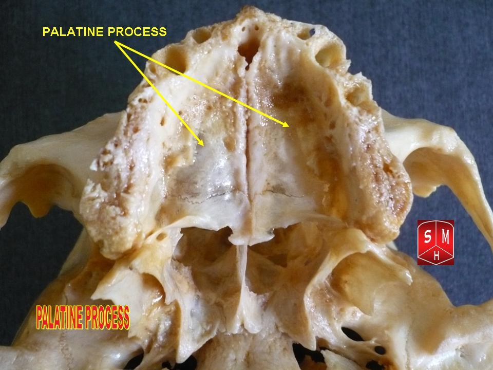
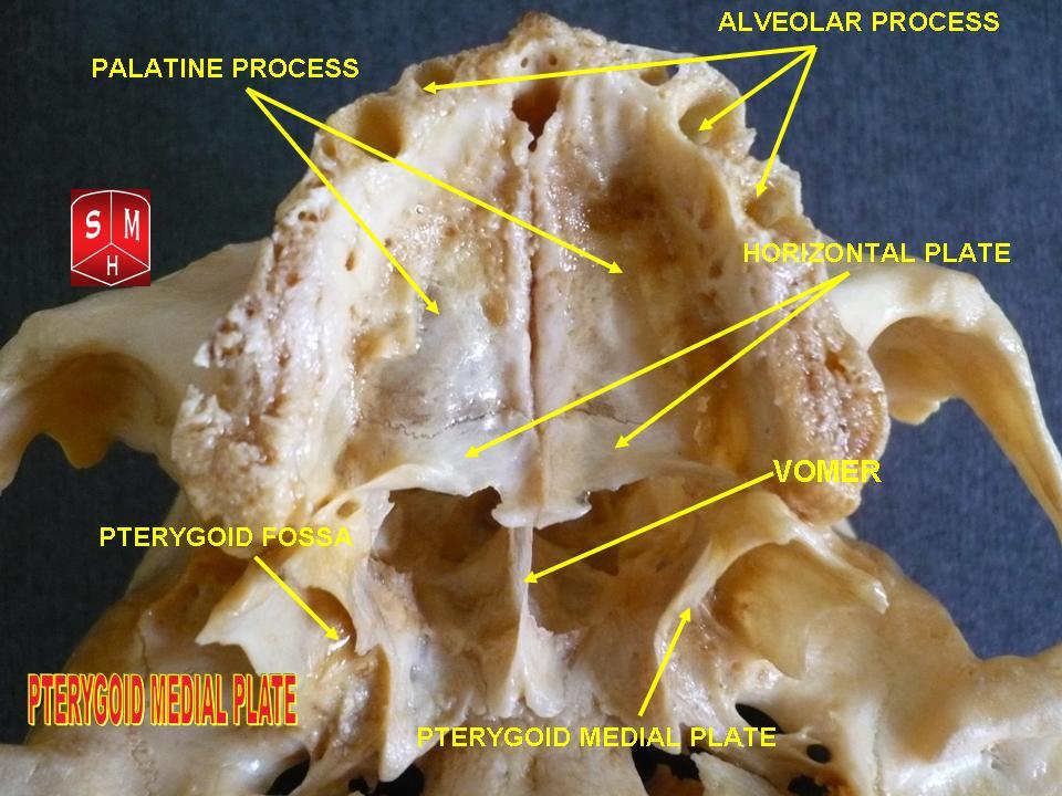
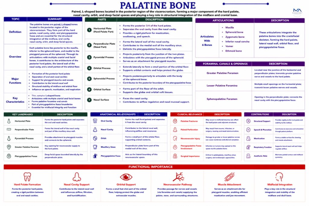
Inferior Surface –  forms the posterior region of the roof of the oral cavity. It is roughened for attachment of the palatal mucosa and connective tissues and contains small openings that transmit branches of the greater palatine vessels and nerves supplying the mucosa of the hard palate.

Superior Surface –  contributes to the floor of the nasal cavity, supporting the respiratory mucosa and participating in the physiological processes of air humidification, warming, and filtration.

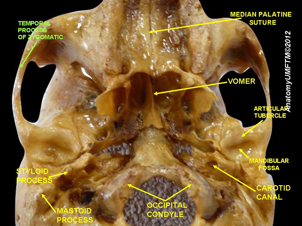
Medial Surface -the nasal cavity and contributes to the posterior portion of the lateral nasal wall, providing articulation sites for structures such as the inferior nasal concha and ethmoid bone.

Lateral Surfaceforms part of the medial boundary of the pterygopalatine fossa, an anatomically complex space that contains branches of the maxillary artery, maxillary nerve (CN V₂), and the pterygopalatine ganglion

Exam Question

Analyze how the different surfaces of the palatine bone contribute to the structural organization and physiological functions of the oral cavity, nasal cavity, and pterygopalatine region, with emphasis on mucosal support and airflow dynamics.

Borders

Anterior Border – articulates with the palatine process of the maxilla, forming the transverse palatine suture, which unites the anterior and posterior components of the hard palate.

Posterior Border forms the posterior margin of the hard palate and provides attachment for the soft palate, which plays an important role in swallowing and speech.

Medial Border articulates with the horizontal plate of the opposite palatine bone, forming the median palatine suture, which maintains symmetry of the posterior palate.

Exam Question

Evaluate how the borders of the palatine bone contribute to the formation and stability of the hard palate, including the structural and functional importance of the transverse and median palatine sutures.

Articulations

Each palatine bone articulates with 6 surrounding bones, forming part of the complex skeletal network of the midface.

These articulations include:

maxilla

sphenoid bone

ethmoid bone

inferior nasal concha

vomer

opposite palatine bone

Through these articulations, the palatine bone connects the nasal cavity, orbit, oral cavity, and cranial base, ensuring the structural stability of the midfacial skeleton.

Exam Question

Discuss how the multiple articulations of the palatine bone with surrounding craniofacial bones contribute to the stability and integration of the midface, and explain their role in maintaining continuity between the nasal, oral, and orbital cavities.

Landmarks

Several clinically important foramina and anatomical features are associated with the palatine bone.

Greater Palatine Foramen– located near the posterior portion of the hard palate, this foramen transmits the greater palatinenerve and vessels, which supply the mucosa and glands of the hard palate.

Lesser Palatine Foraminasituated posterior to the greater palatine foramen, these openings transmit the lesser palatine nerves and vessels, which supply the soft palate and uvula.

Sphenopalatine Foramen – this opening connects the nasal cavity with the pterygopalatine fossa and transmits the sphenopalatine artery, nasopalatine nerve, and posterior nasal nerves.

These neurovascular structures play an essential role in the sensory innervation and vascular supply of the nasal cavity and palate

Exam Question

Explain the anatomical and clinical significance of the greater palatine foramen, lesser palatine foramina, and sphenopalatine foramen, with emphasis on their transmitted neurovascular structures and relevance in surgical and anesthetic procedures.

FUNCTIONAL IMPORTANCE

Oral Seperation

The palatine bones, via their horizontal plates, establish the posterior hard palate, forming a rigid partition between the oral and nasal cavities. 

This separation is essential for coordinated mastication, swallowing, and respiration, preventing food regurgitation into the nasal cavity while allowing simultaneous breathing during feeding

Speech Resonance

By contributing to the hard palate and posterior oral cavity boundaries, the palatine bones provide a stable surface for tongue articulation and phonation. 

They regulate airflow and resonance within oral and nasal chambers, directly influencing speech clarity, articulation of palatal consonants, and overall vocal quality.

Airflow Regulation

Through the perpendicular plate, the palatine bones form part of the lateral nasal wall, shaping posterior nasal airflow dynamics. 

This structural configuration supports air conditioning processes- warming, humidification, and filtration – ensuring efficient respiratory function within the upper airway.

Neurovascular Hub

The palatine bones define the pterygopalatine fossa, a critical neurovascular crossroads of the midface. 

Through foramina and canals, they facilitate transmission of branches of the maxillary nerve (V2), pterygopalatine ganglion fibers, and maxillary artery branches, integrating sensory, autonomic, and vascular supply to the nasal cavity, palate, and orbit.


CLINICAL RELEVANCE

Cleft Pathology

Failure of fusion between the palatine processes during embryological development results in cleft palate, producing persistent communication between oral and nasal cavities. 

This leads to feeding difficulties, nasal regurgitation, hypernasal speech, and abnormal craniofacial growth, requiring multidisciplinary surgical and rehabilitative management

Epistaxis Source

The sphenopalatine artery, passing through the sphenopalatine foramen associated with the palatine bone, represents the principal arterial supply to the nasal cavity. 

Its rupture causes posterior epistaxis, a severe and potentially life-threatening condition often requiring endoscopic ligation or cauterization.

Anesthesia Landmark

The greater palatine foramen, located in the posterior hard palate, serves as a key landmark in dental anesthesia. 

Targeted injection here produces a greater palatine nerve block, enabling painless maxillary procedures such as tooth extraction, periodontal surgery, and palatal grafting

Surgical Corridor

The palatine bone contributes to the boundaries of the pterygopalatine fossa, a region frequently accessed in endoscopic sinus and skull base surgery. 

Precise anatomical knowledge is critical to avoid injury to adjacent neurovascular structures, including the maxillary nerve and arterial branches, during tumor resection or surgical intervention.

SUMMARY TABLE

Scroll to Top