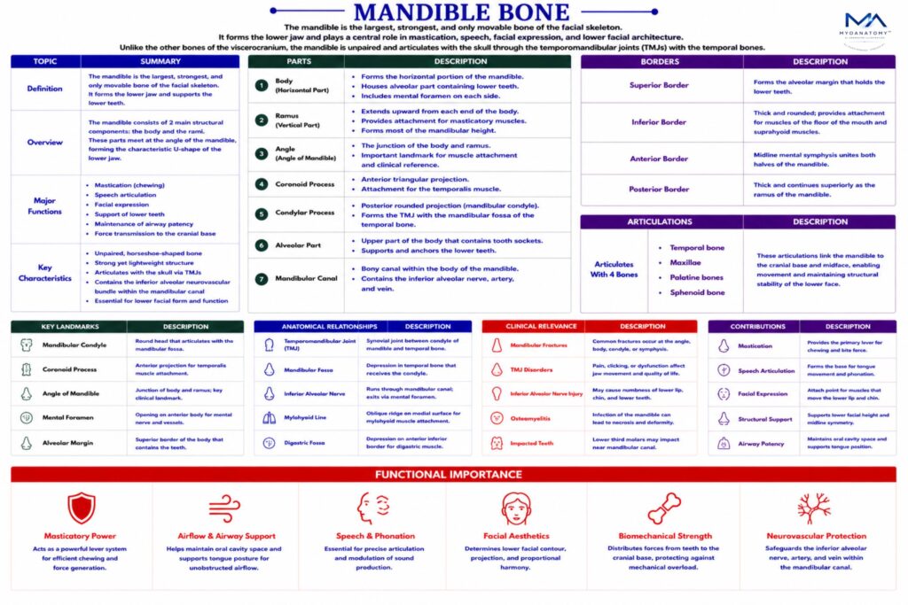
Mandible Bone

The mandible is the largest, strongest, and only movable bone of the facial skeleton. It forms the lower jaw and plays a central role in mastication, speech, facial expression, and lower facial architecture. Unlike the other bones of the viscerocranium, the mandible is unpaired and articulates with the skull through the temporomandibular joints (TMJs) with the temporal bones

CORE

Overview

Structurally, the mandible supports the lower dental arch, provides attachment for numerous muscles of mastication and facial expression, and transmits important neurovascular structures through its canals and foramina.

Because of its functional and biomechanical importance, the mandible is a key element of craniofacial mechanics and oral physiology.

ANATOMY

Parts

The mandible consists of 2 main structural components:

Body of the mandibleforms the  horizontal portion of the mandible and supports the lower teeth.

Ramus of the mandible

These parts meet at the angle of the mandible, forming the characteristic shape of the lower jaw.

Exam Question

Explain how the junction of the mandibular body and ramus at the angle influences force redistribution during mastication and contributes to common fracture patterns.

Surfaces

External surfacecontains several important anatomical features:

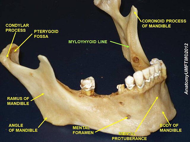
mental protuberance  –  midline prominence forming the chin

mental tubercles  –  small elevations on either side of the chin

mental foramen  –  transmits the mental nerve and vessels, branches of the inferior alveolar neurovascular bundle supplying the lower lip and chin

Internal surface presents important muscular attachments and anatomical landmarks:

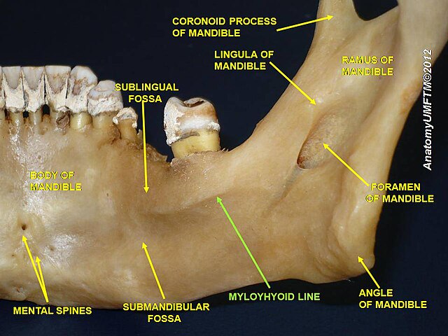
mylohyoid line  –  attachment for the mylohyoid muscle, forming the floor of the oral cavity

digastric fossa  –  attachment for the anterior belly of the digastric muscle

sublingual fossa  –  accommodates the sublingual gland

submandibular fossa  – accommodates the submandibular gland

Lateral surface- provides attachment for the masseter muscle, one of the primary muscles of mastication.

Medial surface– contains the mandibular foramen, which transmits the inferior alveolar nerve and vessels into the mandibular canal. Just inferior to this foramen is the lingula, a small bony projection serving as attachment for the sphenomandibular ligament. Extending anteriorly from the mandibular foramen is the mylohyoid groove, transmitting the mylohyoid nerve and vessels.

Exam Question

Correlate the anatomical features of the medial mandibular surface with the course of the inferior alveolar neurovascular bundle and the functional role of associated muscular attachments.

Process

At the superior end of the ramus are 2  important processes separated by the mandibular notch:

Coronoid Process-  is an anterior triangular projection that serves as the insertion site for the temporalis muscle, a powerful elevator of the mandible.

Condylar Process –  forms the mandibular condyle, which articulates with the mandibular fossa of the temporal bone to form the temporomandibular joint (TMJ). This joint enables movements of the mandible including:

– elevation and depression

– protrusion and retrusion

 -lateral deviation

These movements are essential for mastication and speech.

Exam Question

Critically compare the coronoid and condylar processes in terms of their biomechanical roles, including muscle insertion, force generation, and participation in temporomandibular joint dynamics.

Borders

The mandible presents several anatomical borders:

Inferior border- forms the lower margin of the jaw and provides attachment for muscles of the neck and face.

Posterior border- forms part of the mandibular ramus and contributes to the angle of the mandible.

Anterior border – continuous with the alveolar part of the mandible.

Exam Question

Analyze how the morphology of the mandibular borders reflects patterns of muscle attachment and influences mandibular stability under functional loading

Articulations

The mandible articulates with the skull through the temporomandibular joints, formed between:

mandibular condyle

mandibular fossa of the temporal bone

These joints are synovial joints with an articular disc, allowing complex mandibular movements

Exam Question

Explain how the structural organization of the temporomandibular joint enables synchronized rotational and translational movements, and discuss the consequences of disruption of this coordination.

Landmarks

Important anatomical landmarks of the mandible include:

mental protuberance

mental foramen

mandibular foramen

mandibular canal

mylohyoid line

coronoid process

condylar process

angle of the mandible

These landmarks are important in dentistry, maxillofacial surgery, and anesthetic procedures.

Exam Question

Evaluate the anatomical variability and clinical relevance of key mandibular landmarks, particularly in relation to inferior alveolar nerve block and surgical navigation.

FUNCTIONAL IMPORTANCE

Masticatory Mechanism

The mandible constitutes the primary mobile lever of the craniofacial skeleton, forming the biomechanical foundation of mastication. 

Through articulation at the temporomandibular joint (TMJ), it enables complex multidirectional movements – elevation, depression, protrusion, retrusion, and lateral excursion – generated by coordinated contraction of the muscles of mastication (masseter, temporalis, medial and lateral pterygoids). 

Its alveolar process transmits and distributes occlusal forces, facilitating efficient food comminution while maintaining dynamic equilibrium between bone remodeling and dental loading.


Dental Support

The mandible forms the inferior dental arch, housing the alveoli for the mandibular dentition and providing structural opposition to the maxillary teeth. 

It ensures occlusal alignment and force distribution, essential for mechanical digestion. 

The intimate relationship between tooth roots, alveolar bone, and periodontal structures allows adaptive responses to functional stress, preserving occlusal stability and masticatory efficiency

Muscular Anchorage

The mandible serves as a critical musculoskeletal anchor, providing attachment for both masticatory and suprahyoid muscle groups. 

These include the mylohyoid, geniohyoid, digastric, and genioglossus muscles, which collectively regulate oral floor stability, tongue positioning, and swallowing mechanics. 

Through these attachments, the mandible integrates oropharyngeal coordination, contributing to airway patency, deglutition, and phonatory control

Facial Architecture

The mandible is fundamental to lower facial morphology, determining facial height, contour, and proportional harmony. It provides structural support for soft tissues of the lower face and influences craniofacial biomechanics. 

Its growth and remodeling patterns are essential for maintaining functional occlusion, aesthetic balance, and skeletal integrity, making it a key determinant of both form and function within the craniofacial complex

CLINICAL RELEVANCE

Fracture Mechanic

Mandibular fractures arise from complex vector transmission through a U-shaped cortical–trabecular system, where tensile forces at the superior border and compressive forces at the inferior border interact with muscle pull (masseter, temporalis, pterygoids) to produce predictable displacement and malocclusion patterns.

Neurovascular Injury

Injury to the inferior alveolar nerve within the mandibular canal results from direct fracture line involvement or secondary edema-induced compression, with ischemia and axonal disruption leading to prolonged or permanent sensory deficit due to constrained intrabony healing conditions.

Joint Pathodynamics

Temporomandibular dysfunction reflects failure of coordinated condylar rotation and anterior translation along the articular eminence, where disc displacement and altered synovial loading initiate degenerative remodeling and chronic nociceptive activation

Anesthetic Failure

Variability in mandibular foramen position, lingula morphology, and soft tissue barriers contributes to inferior alveolar nerve block failure, as inaccurate deposition prevents adequate anesthetic diffusion to the nerve prior to its entry into the canal.

SUMMARY TABLE

Scroll to Top