Muscle Anatomy
Muscle anatomy explores the classification, organization, biomechanics, and function of muscles responsible for movement, posture, and joint stability.

Muscle Classification
MYO CORE
Overview
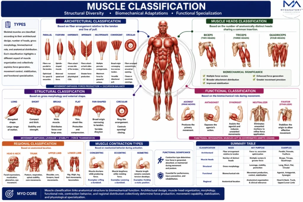
Skeletal muscle classification is a multidimensional anatomical and biomechanical framework that integrates architectural design, muscle-head organization, functional role, regional distribution, and gross morphology. Together, these classification systems explain how structural variations influence force production, excursion, contraction velocity, movement control, stabilization, and functional specialization.
OVERVIEW
Types of Muscle Classification
Skeletal muscle classification is a systematic framework used to organize muscles according to their structural design, anatomical organization, biomechanical properties, and functional roles within the musculoskeletal system.
Because skeletal muscles exhibit remarkable diversity in size, shape, fiber arrangement, attachment patterns, and mechanical behavior, no single classification system can adequately describe all aspects of muscular organization. Instead, multiple complementary classification systems are employed, each emphasizing a distinct anatomical or functional characteristic.
Collectively, these classification methods provide a comprehensive understanding of how muscular structure influences force generation, movement production, joint stabilization, posture maintenance, and functional specialization.
Classification therefore serves not merely as an anatomical cataloging tool, but as a predictive biomechanical model linking morphology to physiological performance.
AI Generated Illustration
Exam Question
How does skeletal muscle classification relate muscle morphology and architectural design to biomechanical function, force generation, movement, and functional specialization?
ANATOMY
Architectural Classification
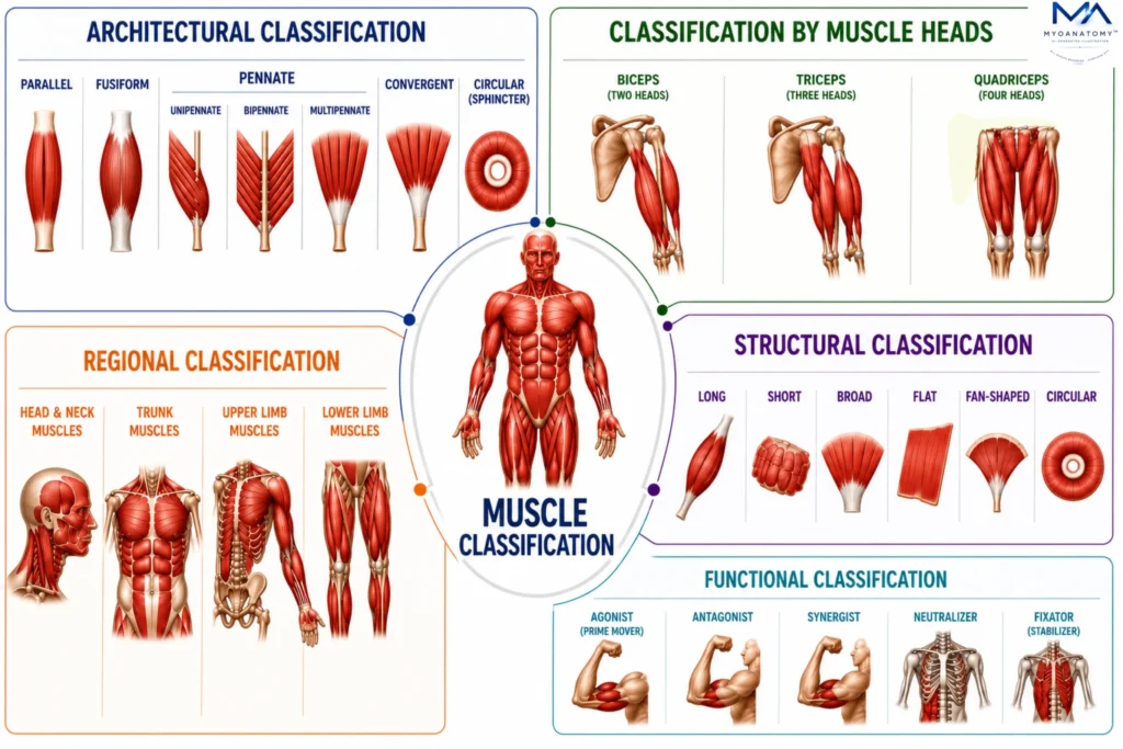
Architectural classification organizes muscles according to the orientation of muscle fibers relative to the tendon and line of pull. It is the most biomechanically significant classification because fiber arrangement directly determines force production, contraction velocity, excursion, and mechanical efficiency.
The relationship between fiber length and physiological cross-sectional area (PCSA) dictates whether a muscle is optimized for rapid movement, extensive range of motion, stabilization, or maximal force generation.
Consequently, muscle architecture serves as the strongest anatomical predictor of functional performance.
Representative examples include the parallelly arranged sartorius, the fusiform biceps brachii, the unipennate extensor digitorum longus, the bipennate rectus femoris, the multipennate deltoid muscle, the convergent pectoralis major, and the circular orbicularis oris.
Exam Question
How does muscle fiber architecture influence force production, contraction velocity, excursion, and mechanical efficiency, and why do muscles such as the sartorius, deltoid, and pectoralis major exhibit distinct biomechanical capabilities despite sharing the same fundamental contractile mechanism?
Muscle Heads Classification
Muscle-head classification categorizes muscles according to the number of anatomically distinct heads that converge into a common tendon or insertion.
Each head contributes a unique line of pull and mechanical vector, increasing structural complexity and functional versatility.
By integrating forces from multiple origins, multi-headed muscles enhance force distribution, movement precision, joint stabilization, and biomechanical efficiency across a wider range of motion.
This classification is exemplified by the two-headed biceps brachii, the three-headed triceps brachii, and the four-headed quadriceps femoris.
Exam Question
How does increasing the number of muscular heads alter force distribution, movement versatility, and joint stabilization, and what functional advantages distinguish the biceps brachii, triceps brachii, and quadriceps femoris?
Structural Classification
Structural classification organizes muscles according to their overall shape and geometric configuration.
Unlike architectural classification, which focuses on fiber orientation, this system emphasizes external form and anatomical appearance.
Variations in muscular morphology reflect adaptations to regional anatomy, attachment patterns, and mechanical demands, influencing force transmission, leverage, stability, and movement capability.
Examples include the long sartorius, the short interossei muscles, the broad latissimus dorsi, the flat external oblique, the fan-shaped temporalis, and the circular orbicularis oculi.
Exam Question
How do differences in muscular shape and gross morphology influence force transmission, leverage, and mechanical function, and how are these adaptations reflected in muscles such as the sartorius, temporalis, latissimus dorsi, and external oblique?
Functional Classification
Functional classification categorizes muscles according to their biomechanical role during movement.
Rather than acting independently, muscles function within coordinated kinetic chains where specific muscles generate movement, oppose motion, stabilize joints, refine force vectors, or eliminate unwanted actions.
This classification highlights the neuromuscular coordination required for efficient movement, postural control, mechanical precision, and joint integrity.
For example, during elbow flexion the biceps brachii acts as an agonist, the triceps brachii functions as an antagonist, the brachialis serves as a synergist, the pronator teres may act as a neutralizer, and the trapezius functions as a fixator by stabilizing the shoulder girdle.
Exam Question
How do agonists, antagonists, synergists, neutralizers, and fixators interact during coordinated movement, and how do muscles such as the biceps brachii, triceps brachii, brachialis, pronator teres, and trapezius collectively contribute to efficient elbow flexion and upper-limb stability?
Regional Classification
Regional classification organizes muscles according to their location within specific anatomical regions of the body.
Muscles within the same region often share common neurovascular supply, fascial compartments, developmental origins, and functional relationships. This classification provides a systematic framework for anatomical study, clinical localization, radiological interpretation, and understanding coordinated regional movement patterns.
Representative examples include the masseter within the head and neck region, the rectus abdominis within the trunk, the biceps brachii within the upper limb, and the gluteus maximus within the lower limb.
Exam Question
How does regional organization of skeletal muscles facilitate anatomical specialization and coordinated function, and how do the masseter, rectus abdominis, biceps brachii, and gluteus maximus exemplify the distinct biomechanical demands of their respective body regions?
SUMMARY TABLE