
Muscle
Clinical Relevance
MYO CORE

Overview
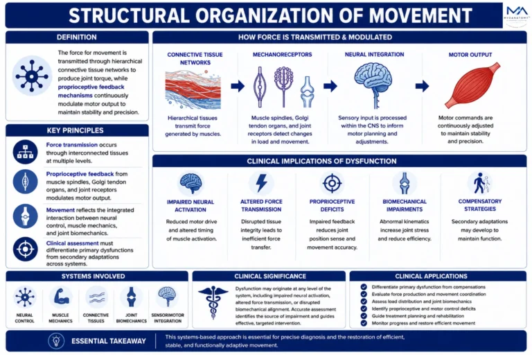
Clinical evaluation of the musculoskeletal system requires interpretation of movement as the integrated output of a multiscale neuromuscular–biomechanical system. Skeletal muscle function is governed by α-motor neuron activation, motor unit recruitment dynamics, and excitation–contraction coupling, which together regulate force generation at the sarcomere level.
OVERVIEW
This force is transmitted through hierarchical connective tissue networks to produce joint torque, while proprioceptive feedback mechanisms – mediated by muscle spindles, Golgi tendon organs, and joint receptors – continuously modulate motor output to maintain stability and precision. As a result, observable movement reflects the coordinated interaction between neural control, muscle mechanics, and joint biomechanics.
Clinically, dysfunction may originate at any level of this system, including impaired neural activation, altered force transmission, or disrupted biomechanical alignment. However, such deficits rarely present in isolation; instead, they manifest as integrated movement impairments characterized by altered kinematics, reduced force efficiency, and compensatory motor strategies.
Accurate clinical assessment therefore requires differentiation between primary dysfunction and secondary adaptations, with evaluation of force production, movement coordination, and load distribution across the system. This systems-based approach is essential for precise diagnosis and the restoration of efficient, stable, and functionally adaptive movement.
AI generated Illustration MyoAnatomy.
Exam Question
How do neuromuscular control, force transmission, and joint biomechanics interact to produce normal movement, and how does dysfunction alter this system?
CLINICAL EVALUATION
Range of Motion
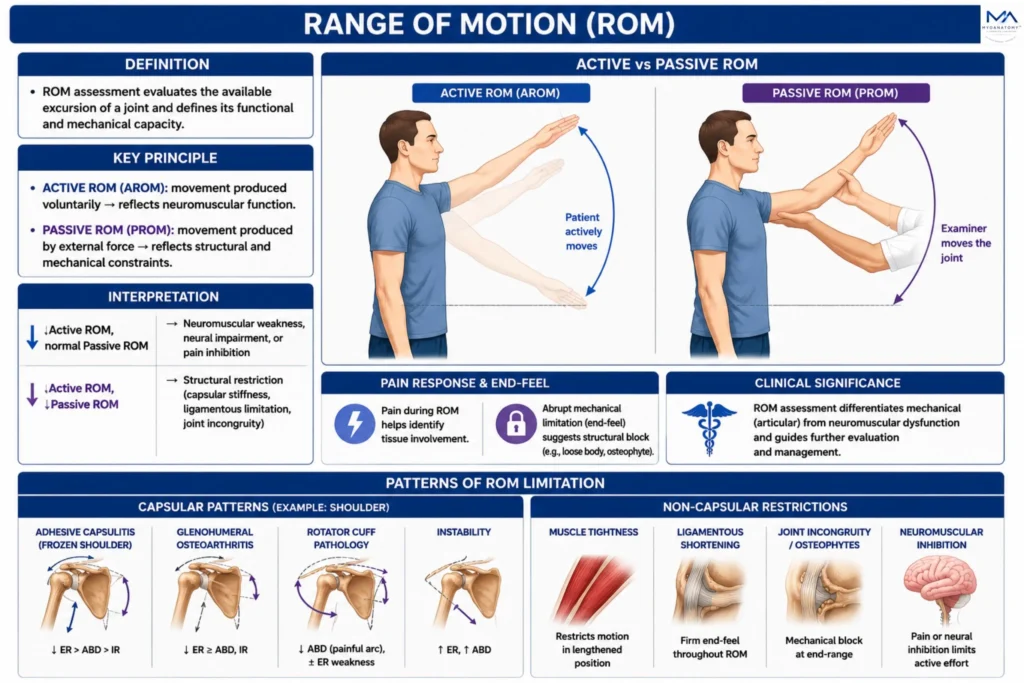
Range of motion (ROM) assessment evaluates the available excursion of a joint and thereby defines its functional and mechanical capacity. By comparing active ROM, which reflects voluntary neuromuscular performance, with passive ROM, which reflects the integrity and extensibility of articular and periarticular structures, ROM analysis helps localize the source of dysfunction.
Limitation of active ROM with preserved passive motion suggests impairment of the contractile or neural apparatus, whereas concurrent restriction of both active and passive ROM indicates structural limitation within the joint or surrounding soft tissues.
In addition, ROM assessment identifies capsular and non-capsular patterns of restriction, characterizes pain behavior during movement, and may reveal abrupt mechanical block suggestive of intra-articular obstruction.
Clinically, it is a fundamental tool for distinguishing mechanical articular dysfunction from neuromuscular impairment and for directing further musculoskeletal evaluation.
AI generated Illustration MyoAnatomy.
Exam Question
How does comparison of active and passive range of motion distinguish neuromuscular dysfunction from structural joint restriction, and what can patterns of limitation and pain response reveal about the underlying pathology?
Manual Muscle Testing
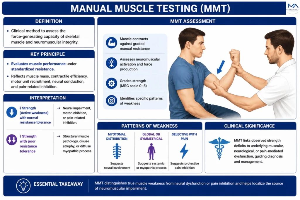
Manual Muscle Testing (MMT) is a clinical method used to assess the force-generating capacity of skeletal muscle and the functional integrity of the neuromuscular system.
It evaluates muscle performance under standardized resistance and reflects the combined influence of muscle mass, contractile efficiency, motor unit recruitment, neural conduction, and pain-related inhibition.
Reduction in muscle strength may result from impaired neural activation, defective excitation–contraction coupling, structural muscle pathology, or disuse-related atrophy. The distribution of weakness is diagnostically important: myotomal weakness suggests neurogenic involvement, diffuse or symmetrical weakness may indicate systemic or myopathic disease, and selective painful weakness often reflects protective inhibition rather than primary loss of contractile function.
Clinically, MMT provides both functional and diagnostic information by linking observed strength deficits to underlying muscular, neural, or pain-mediated dysfunction.
AI generated Illustration MyoAnatomy.
Exam Question
How does manual muscle testing differentiate true muscle weakness from neural dysfunction or pain-mediated inhibition, and what is the diagnostic significance of specific weakness patterns?
Gait Analysis
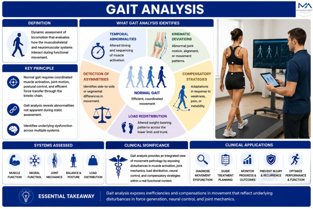
Gait analysis is the dynamic assessment of locomotion and evaluates how the musculoskeletal and neuromuscular systems interact during functional movement.
Because normal gait depends on precisely coordinated muscle activation, joint motion, postural control, and efficient transfer of forces through the kinetic chain, gait examination reveals abnormalities that are not apparent during static assessment.
It allows identification of temporal disturbances in movement sequencing, kinematic deviations in joint alignment and excursion, compensatory strategies adopted in response to pain, weakness, or instability, and altered load distribution across the lower limb and axial skeleton.
Clinically, gait analysis provides an integrated view of movement pathology by exposing disturbances in force generation, neural control, balance, and joint mechanics within a real functional context.
AI generated Illustration MyoAnatomy.
Exam Question
How does gait analysis reveal abnormalities in movement coordination, joint mechanics, and load distribution, and why are these findings important in identifying functional movement pathology?
Neurological Assessment
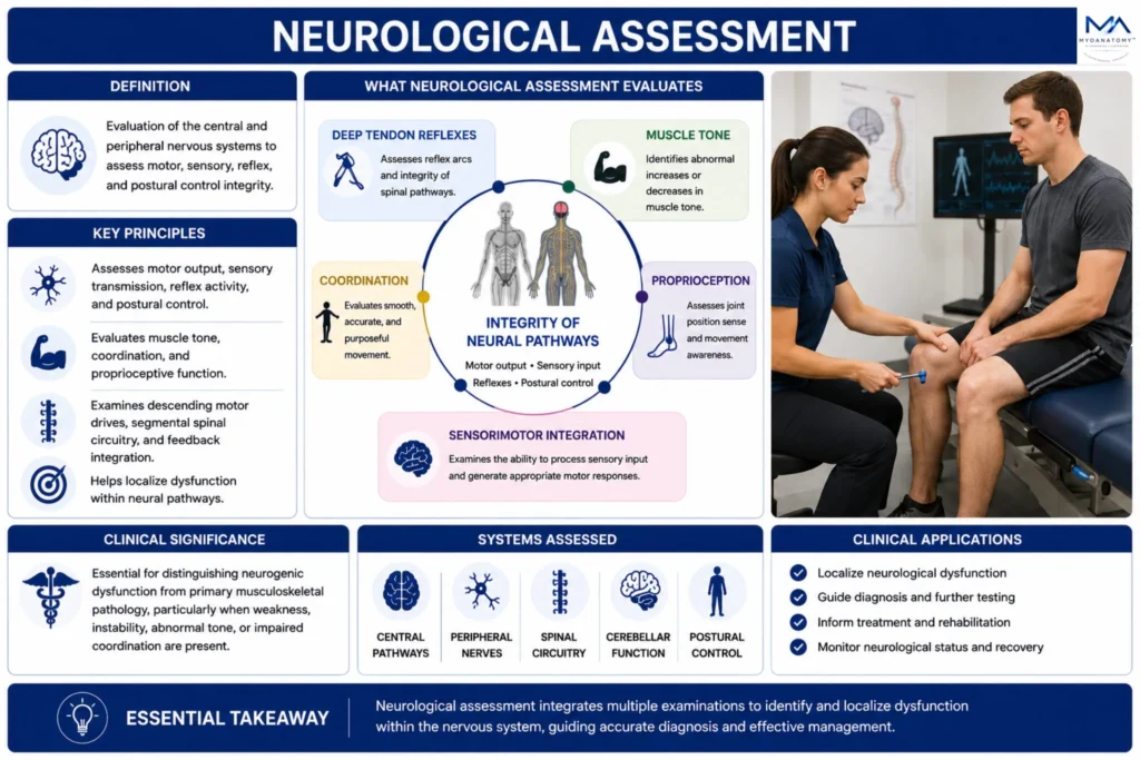
Neurological examination assesses the integrity of the central and peripheral pathways responsible for motor output, sensory transmission, reflex activity, and postural control.
Through evaluation of deep tendon reflexes, muscle tone, coordination, and proprioceptive function, it examines the adequacy of descending motor drive, segmental spinal circuitry, afferent feedback, and sensorimotor integration.
Abnormal findings such as hyperreflexia, spasticity, flaccidity, ataxia, or impaired joint position sense indicate disruption of neural regulation and may localize pathology to upper motor neuron, lower motor neuron, cerebellar, or sensory pathways.
Clinically, neurological assessment is essential for distinguishing neurogenic dysfunction from primary musculoskeletal pathology, particularly in the presence of weakness, instability, abnormal tone, or impaired coordination.
AI generated Illustration MyoAnatomy.
Exam Question
How do reflexes, muscle tone, coordination, and proprioception collectively assess neural pathway integrity and help distinguish neurological dysfunction from primary musculoskeletal pathology?