Muscle
Structural Organization
MYO CORE
Muscle Fiber Organization
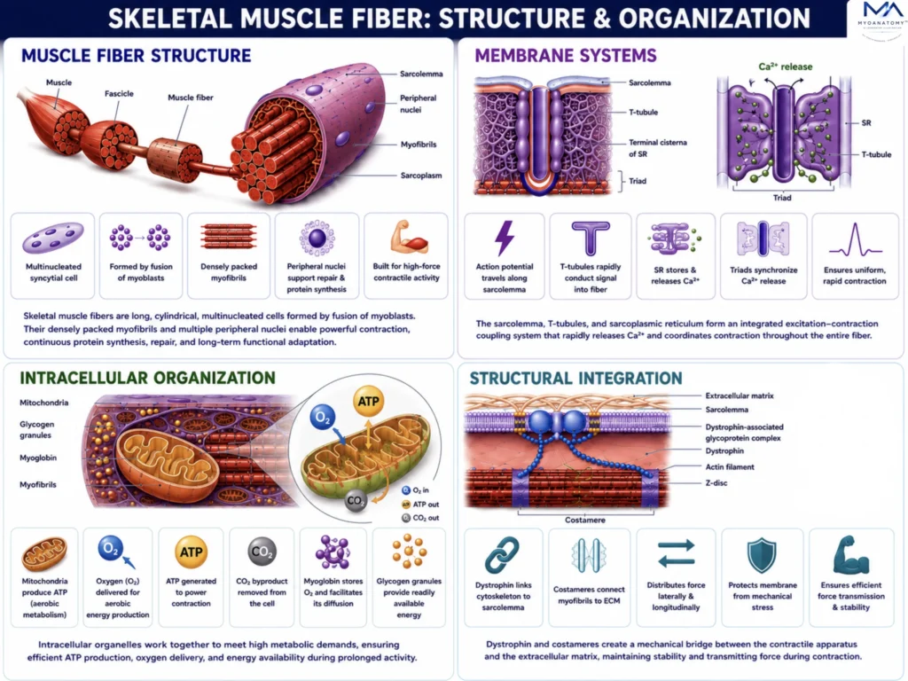
Skeletal muscle fibers are highly specialized multinucleated cells that integrate membrane systems, metabolic organelles, contractile proteins, and structural support networks into a unified force-producing unit. Coordinated excitation–contraction coupling, efficient ATP generation, and specialized force-transmission mechanisms enable rapid, synchronized contraction while maintaining cellular stability and mechanical efficiency.
OVERVIEW
Skeletal muscle fibers possess a highly specialized organization that integrates electrical activation, intracellular metabolism, contractile function, and structural stability into a single coordinated system. This hierarchical arrangement ensures efficient conversion of neural signals into controlled force production.
The sarcolemma, T-tubules, and sarcoplasmic reticulum form an integrated excitation–contraction coupling network that rapidly propagates action potentials and synchronizes calcium release throughout the fiber. This mechanism enables simultaneous activation of myofibrils and precise regulation of contraction.
Within the sarcoplasm, mitochondria, glycogen granules, and myoglobin provide the metabolic foundation required for sustained ATP production, oxygen utilization, and continuous muscular activity. Their strategic distribution supports rapid energy delivery directly to active contractile sites.
Mechanical integrity is maintained by specialized cytoskeletal proteins, including dystrophin and costameres, which connect sarcomeres to the sarcolemma and extracellular matrix. These structures stabilize the muscle fiber, distribute contractile stresses, and ensure efficient transmission of force from the molecular level to the entire muscle.
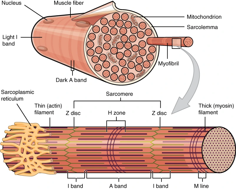
Organization of Muscle Fiber – OpenStax Anatomy & Physiology, Wikimedia Commons. Creative Commons Attribution 4.0 International (CC BY 4.0).
Exam Question
Evaluate how skeletal muscle fiber organization coordinates electrical signaling, calcium regulation, metabolic support, and cytoskeletal force transmission to ensure efficient contraction and long-term maintenance of muscular function.
ANATOMY
Muscle Fiber Structure
A skeletal muscle fiber is a highly specialized multinucleated syncytium optimized for sustained force production and mechanical efficiency.
Formed through the fusion of embryonic myoblasts, each fiber contains numerous peripheral nuclei that collectively regulate protein synthesis, cellular maintenance, and adaptive remodeling. The sarcoplasm is densely occupied by parallel myofibrils, maximizing the volume dedicated to contraction while minimizing non-contractile space.
This structural arrangement enables the fiber to generate substantial force, respond to mechanical loading, and maintain functional integrity through continuous cycles of contraction, repair, and adaptation.
Exam Question
How do the multinucleated organization and myofibrillar architecture of skeletal muscle fibers contribute to force generation, cellular maintenance, and adaptive plasticity?
Membrane Systems
The sarcolemma, T-tubules, and sarcoplasmic reticulum form an integrated membrane network responsible for excitation–contraction coupling.
Action potentials propagate along the sarcolemma and rapidly penetrate the fiber through the T-tubule system, ensuring that electrical activation reaches deep contractile regions. Depolarization triggers calcium release from the terminal cisternae of the sarcoplasmic reticulum, initiating actin–myosin interaction throughout the fiber.
This highly specialized arrangement synchronizes myofibrillar activation and enables rapid, uniform, and precisely controlled contraction regardless of fiber size.
Exam Question
Explain how the structural organization of the sarcolemma, T-tubules, and sarcoplasmic reticulum ensures efficient excitation-contraction coupling in skeletal muscle.
Intracellular Organization
The sarcoplasm is organized to support the exceptionally high metabolic demands of muscle contraction.
Mitochondria are strategically positioned near myofibrils to provide localized ATP production for cross-bridge cycling and calcium transport. Glycogen granules serve as readily available energy reserves, while myoglobin facilitates intracellular oxygen storage and diffusion.
The close spatial relationship between energy-producing organelles and contractile structures minimizes diffusion constraints, enhances metabolic efficiency, and supports sustained muscular performance during both aerobic and anaerobic activity.
Exam Question
Evaluate how the intracellular organization of skeletal muscle fibers optimizes energy production, oxygen utilization, and resistance to fatigue.
Structural Integration
Efficient muscle function requires not only force generation but also effective force transmission and mechanical stability.
Dystrophin forms a critical link between the intracellular actin cytoskeleton and the dystrophin-associated glycoprotein complex within the sarcolemma, protecting the membrane from contraction-induced stress.
Costameres align with Z-discs and mechanically couple myofibrils to the extracellular matrix, distributing contractile forces both longitudinally and laterally across the fiber. Together, these structures preserve membrane integrity, minimize mechanical injury, and ensure that sarcomeric force is efficiently transmitted to connective tissues and ultimately to the skeleton.
Exam Question
Critically discuss the roles of dystrophin and costameres in maintaining sarcolemmal stability and mediating force transmission during skeletal muscle contraction.
SUMMARY TABLE