Muscle
Structural Organization
MYO CORE
Myofibril Organization
Myofibrils form the fundamental biomechanical framework of skeletal muscle, where serially aligned sarcomeres integrate force generation, force transmission, and structural stability into a single contractile system. This highly ordered architecture synchronizes molecular contractile activity, preserves mechanical continuity, and enables efficient propagation of force throughout the muscle fiber, providing the structural basis for muscle contraction.
OVERVIEW
Myofibril organization represents the fundamental biomechanical framework of skeletal muscle, integrating contractile, elastic, and cytoskeletal elements into a highly ordered force-transmission system.
This architecture ensures that forces generated by actin–myosin cross-bridge cycling are synchronized across thousands of sarcomeres and efficiently propagated throughout the muscle fiber.
Beyond force production, myofibrillar organization preserves sarcomeric alignment, maintains mechanical continuity, and protects the contractile apparatus from deformation during repetitive loading.
Through the coordinated interaction of contractile and structural proteins, myofibrils function as dynamic mechanical networks that optimize force transmission, contractile efficiency, and cellular stability.

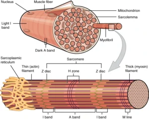
“Organization of Muscle Fiber” – OpenStax Anatomy & Physiology via Wikimedia Commons. Licensed under CC BY 4.0
Exam Question
Analyze how the structural organization of myofibrils integrates contractile, elastic, and cytoskeletal elements to optimize force transmission, mechanical stability, and skeletal muscle performance.”
ANATOMY
Myofibril Assembly
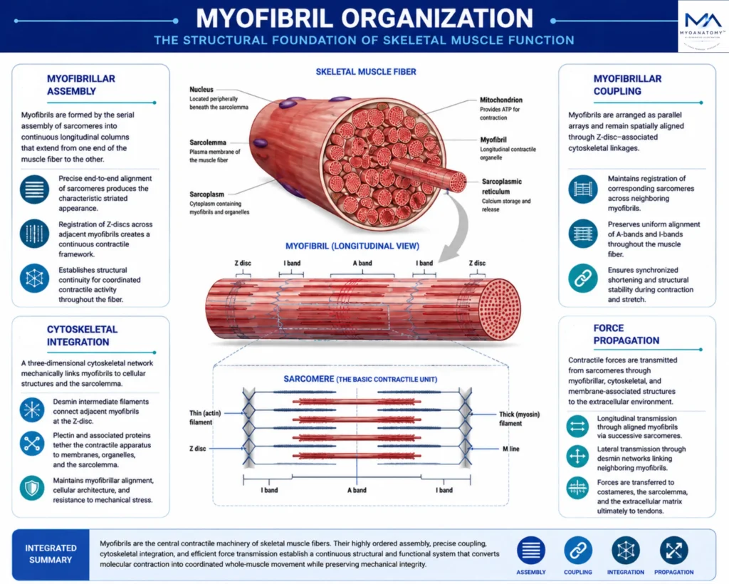
Myofibrils are highly organized contractile organelles that occupy the majority of the sarcoplasm and extend continuously throughout the length of the muscle fiber.
They are formed by the serial assembly of sarcomeres into longitudinal contractile columns whose precise alignment produces the characteristic cross-striated appearance of skeletal muscle.
Registration of Z-discs across adjacent myofibrils establishes a continuous intracellular framework that preserves architectural organization and enables coordinated contractile activity throughout the fiber.
Exam Question
Critically evaluate how the hierarchical assembly of myofibrils from serially aligned sarcomeres establishes the structural foundation for coordinated force generation, intracellular organization, and skeletal muscle contraction.
Myofibril Coupling
Myofibrils function as components of an integrated contractile network rather than as isolated structures.
Their spatial alignment is maintained through Z-disc-associated cytoskeletal linkages that preserve the registration of corresponding sarcomeres across neighboring myofibrils.
This organization maintains uniform alignment of contractile bands, stabilizes the myofibrillar lattice during contraction, and ensures synchronized shortening throughout the muscle fiber.
Exam Question
Discuss how myofibrillar coupling contributes to sarcomeric registration, maintenance of the striated architecture, and synchronization of contractile activity throughout skeletal muscle fibers.
Cytoskeletal Integration
The myofibrillar apparatus is mechanically integrated into the cellular architecture through a complex cytoskeletal network centered on the Z-disc.
Desmin intermediate filaments interconnect adjacent myofibrils, while plectin and associated scaffolding proteins anchor the contractile system to membrane complexes, organelles, and the sarcolemma.
This three-dimensional framework preserves sarcomeric alignment, maintains intracellular organization, and provides structural resilience against repetitive mechanical stress.
Exam Question
Analyze the anatomical and functional roles of desmin, plectin, and associated cytoskeletal networks in maintaining myofibrillar alignment, cellular architecture, and resistance to mechanical stress.
Force Propagation
Myofibrils form the principal intracellular pathway for force transmission within skeletal muscle fibers.
Mechanical forces generated by sarcomeric contraction are propagated longitudinally through successive contractile units and distributed laterally through cytoskeletal networks linking adjacent myofibrils.
These forces are subsequently transferred to costameres, the sarcolemma, and the extracellular matrix, creating a continuous anatomical continuum that connects molecular force generation to whole-muscle movement while maintaining mechanical integrity and load distribution.
Exam Question
Critically discuss the pathways through which contractile forces are transmitted from sarcomeres through myofibrillar, cytoskeletal, and membrane-associated structures to generate coordinated whole-muscle movement.
SUMMARY TABLE