The orbicularis oculi is a circular muscle of facial expression that surrounds the orbital opening and eyelids, functioning as the primary sphincter of the palpebral fissure. It is responsible for closure of the eyelids, blinking, squinting, and protection of the eye.

Orbicularis Oculi Muscle

CORE

Overview

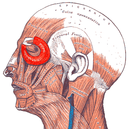
Unlike most skeletal muscles that attach from bone to bone, the orbicularis oculi originates from bony margins of the orbit and adjacent ligaments but inserts largely into the skin and connective tissue of the eyelids and surrounding facial integument, enabling direct movement of the eyelid structures.

Anatomically, the muscle is subdivided into three functional parts, each with distinct roles:

orbital part (pars orbitalis) – responsible for forceful eyelid closure

palpebral part (pars palpebralis) – responsible for gentle blinking

lacrimal part (pars lacrimalis or Horner’s muscle) – assists in tear drainage

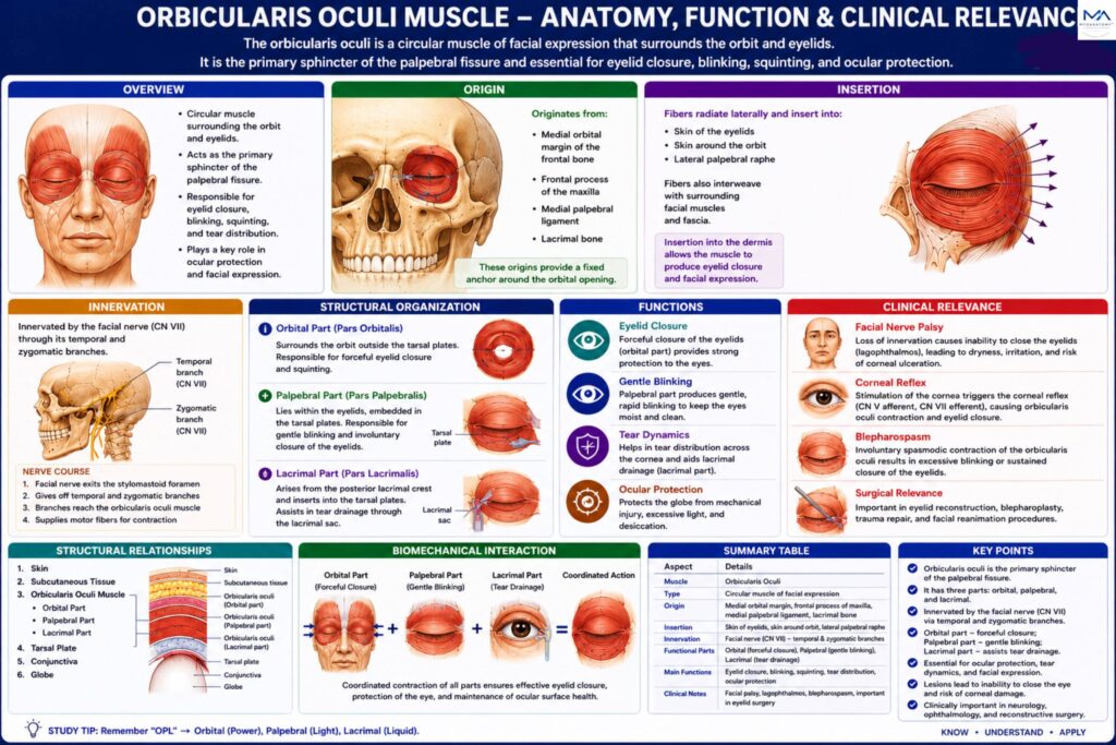
The orbicularis oculi is the principal muscle responsible for closure of the eyelids and protection of the eye. Through its orbital, palpebral, and lacrimal components, it performs multiple functions including blinking, tear distribution, and drainage of lacrimal fluid.

Its close integration with the facial nerve, lacrimal apparatus, and ocular reflex pathways

ANATOMY

Origin

The orbicularis oculi originates from structures located on the medial margin of the orbit, including:

medial orbital margin of the frontal bone

frontal process of the maxilla

medial palpebral ligament

lacrimal bone

These origins provide a fixed anchor from which the circular fibers can surround the orbital opening.

Exam Question

Describe the anatomical origin of the orbicularis oculi muscle and explain how its medial orbital attachments contribute to eyelid function and orbital mechanics.

Insertion

The muscle fibers radiate laterally and insert into:

skin of the eyelids

skin surrounding the orbit

lateral palpebral raphe

Because the muscle inserts into the dermis of the eyelids, contraction directly alters eyelid position.

Exam Question

Explain the insertion pattern of the orbicularis oculi and discuss why insertion into the eyelids and surrounding facial integument is functionally important compared with typical skeletal muscles.

Structural Organization

Orbital Part –  orbital portion forms the outermost circular fibers surrounding the orbit.

characteristics:

thick muscular fibers; extend beyond orbital margins

responsible for forceful closure of the eyelids

this portion is activated during:

bright light exposure; protective reflexes; voluntary eye closure.

Palpebral Part –  palpebral portion lies within the eyelids themselves.

characteristics:

thin muscle fibers

extend between the medial and lateral palpebral ligaments.

This part produces gentle closure of the eyelids, as occurs during:

normal blinking; sleep.

Blinking spreads the tear film across the cornea, maintaining ocular lubrication.

Lacrimal Part (Horner’s Muscle)-  arises from the posterior lacrimal crest of the lacrimal bone and inserts into the tarsal plates of the eyelids.

this component:

pulls the eyelids medially; compresses the lacrimal sac

assists tear drainage through the nasolacrimal duct.

Exam Question

Describe the orbital, palpebral, and lacrimal parts of the orbicularis oculi and compare their anatomical arrangement and specialized functional roles.

Innervation

The orbicularis oculi receives motor innervation from the facial nerve (CN VII).

Major branches include:

temporal branch of facial nerve

zygomatic branch of facial nerve

These branches form a neural network across the upper face that controls eyelid movement.

Exam Question

Explain the motor innervation of the orbicularis oculi, including the facial nerve branches involved, and discuss the functional consequences of facial nerve injury.

FUNCTIONAL ROLE

Eyelid Closure

The orbicularis oculi is the principal sphincter of the eyelids, responsible for closure of the palpebral fissure. 

The palpebral part mediates gentle blinking, whereas the orbital part produces forceful eyelid closure during protective responses and voluntary eye squeezing.

Exam Question

How do the orbital and palpebral parts of the orbicularis oculi cooperate to produce both gentle blinking and forceful eyelid closure?

Occular Protection

By closing the eyelids, the orbicularis oculi protects the globe from mechanical injury, excessive light exposure, and foreign particles. 

It forms the efferent component of the corneal reflex, where corneal stimulation (CN V₁) triggers rapid eyelid closure through the facial nerve (CN VII).

Exam Question

Explain the role of the orbicularis oculi in ocular protection and describe its contribution to the corneal reflex pathway.

Tear Dynamics

Repeated blinking distributes the tear film uniformly across the corneal surface, ensuring lubrication, debris removal, and maintenance of optical clarity. Simultaneously, the lacrimal part facilitates tear drainage by compressing the lacrimal sac and directing tears into the nasolacrimal drainage pathway.

Exam Question

Describe how the orbicularis oculi contributes to tear distribution and lacrimal drainage during normal blinking.

Ocular Integration

The orbicularis oculi functions within an integrated protective system involving the eyelids, lacrimal apparatus, extraocular muscles, and facial musculature. 

Through coordinated activity with the levator palpebrae superioris, it enables controlled eyelid movements and supports one of the most frequently performed motor actions in the body- blinking

Exam Question

Discuss how the orbicularis oculi functions within the integrated eyelid–lacrimal–ocular system to maintain visual function and ocular surface health.

CLINICAL RELEVANCE

Facial Paralysis

Damage to the facial nerve (CN VII) results in paralysis of the orbicularis oculi and loss of effective eyelid closure. Patients develop lagophthalmos (inability to close the eye completely), reduced blinking, and impaired corneal protection. Prolonged exposure of the cornea may lead to dryness, irritation, ulceration, and exposure keratopathy. 

Bell’s phenomenon, characterized by upward rotation of the eyeball during attempted eyelid closure, is commonly observed

Exam Question

Explain the clinical manifestations of orbicularis oculi paralysis following facial nerve (CN VII) injury and discuss the mechanisms underlying lagophthalmos and exposure keratopathy.

Cornea Reflex

The orbicularis oculi forms the motor (efferent) component of the corneal reflex. Stimulation of the cornea normally triggers rapid bilateral eyelid closure to protect the eye from injury. 

Facial nerve lesions abolish this protective response despite intact corneal sensation, making corneal reflex testing an important component of neurological examination.

Exam Question

Why is assessment of the corneal reflex clinically important, and how does orbicularis oculi dysfunction help localize neurological lesions?

Blepharospasm

Blepharospasm is a disorder characterized by involuntary spasmodic contraction of the orbicularis oculi. Patients experience excessive blinking or sustained eyelid closure that may interfere with vision and daily activities. 

The condition is often associated with abnormalities of facial motor control and central nervous system regulation.

Exam Question

Describe the anatomical basis of blepharospasm and explain how abnormal orbicularis oculi activity can interfere with normal visual function.

Surgical Relevance

Detailed understanding of orbicularis oculi anatomy is essential during eyelid, orbital, and reconstructive surgery. Procedures such as blepharoplasty, trauma reconstruction, and periocular surgery require preservation of muscular function to maintain normal blinking, effective eyelid closure, corneal protection, and proper lacrimal drainage. 

Improper surgical management may result in functional and cosmetic complications.

Exam Question

Why is detailed knowledge of orbicularis oculi anatomy important in ophthalmic, oculoplastic, and facial reconstructive surgery?

SUMMARY TABLE

Scroll to Top