The buccinator is a broad, thin quadrilateral muscle forming the muscular foundation of the cheek and an essential component of the oral musculature involved in mastication, speech, and maintenance of the oral vestibule. Although classified among the muscles of facial expression, functionally it also plays a critical role in mastication, working closely with the muscles of mastication and the orbicularis oris.
Buccinator Muscle
CORE
Overview
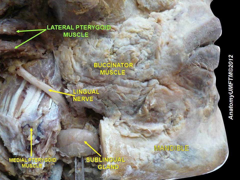
The muscle lies deep within the cheek, forming the lateral wall of the oral cavity, specifically the oral vestibule, the space between the teeth and the cheeks. It is covered
Externally by the buccal fat pad and facial fascia, while internally it is lined by the buccal mucosa.
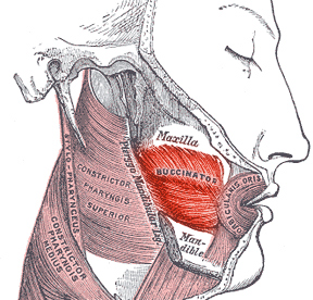
Posteriorly, the buccinator attaches to the pterygomandibular raphe, a fibrous band that connects the muscle to the superior pharyngeal constrictor, illustrating the anatomical continuity between the oral cavity and the pharynx.
Anteriorly, its fibers converge toward the modiolus, where they interlace with the fibers of the orbicularis oris and several other perioral muscles.
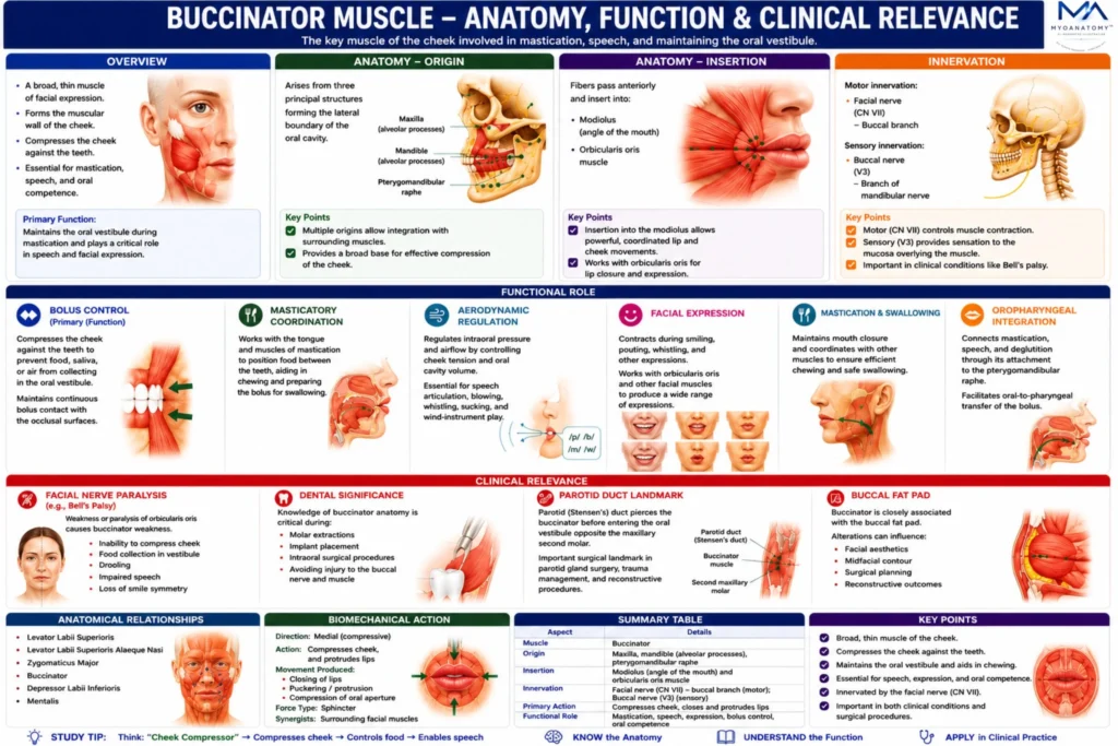
The buccinator muscle forms the muscular framework of the cheek and plays a crucial role in mastication, speech, and maintenance of the oral vestibule. By compressing the cheek against the teeth, it ensures efficient chewing and prevents food from accumulating in the vestibule of the mouth.
Functionally integrated with the orbicularis oris, muscles of mastication, and pharyngeal musculature, the buccinator represents an essential link between the facial expression muscles and the oral cavity’s functional mechanics.
Clinically, its significance is highlighted in facial nerve paralysis, dental procedures, and parotid duct anatomy, making it an important structure in both anatomical and surgical contexts.


ANATOMY
Origin
The buccinator arises from three principal structures forming the lateral boundary of the oral cavity:
alveolar processes of the maxilla opposite the maxillary molar teeth
alveolar processes of the mandible opposite the mandibular molar teeth
pterygomandibular raphe, a fibrous band extending from the pterygoid hamulus of the sphenoidbone to the posterior end of the mylohyoid line of the mandible
This raphe serves as a tendinous attachment shared with the superior pharyngeal constrictor muscle.
Exam Question
Describe the anatomical origins of the buccinator muscle, including its attachments to the maxillary and mandibular alveolar processes and the pterygomandibular raphe, and explain the functional importance of each attachment site.
Insertion
The fibers of the buccinator pass anteriorly and insert into:
modiolus (angle of the mouth)
orbicularis oris muscle
Many fibers interlace with fibers of the orbicularis oris, allowing coordinated control of the lips and cheeks.
Exam Question
Explain the insertion of the buccinator into the modiolus and orbicularis oris, and discuss how this arrangement contributes to coordinated movements of the lips, cheeks, and oral commissure during mastication and facial expression.
Innervation
Motor innervation is provided by the:
buccal branch of the facial nerve (CN VII)
It is important to note that the buccal nerve of the mandibular division of the trigeminal nerve (V3) passes superficially to the buccinator but provides sensory innervation only, not motor supply.
Exam Question
Differentiate the motor and sensory innervation associated with the buccinator muscle, identifying the role of the buccal branch of the facial nerve (CN VII) and the buccal nerve of the mandibular division of the trigeminal nerve (V3). Why is this distinction clinically important.
FUNCTIONAL ROLE
Bolus Control
The buccinator serves as the principal muscular regulator of the oral vestibule, compressing the cheek against the dental arches to prevent sequestration of food between the teeth and cheek.
This action maintains continuous bolus contact with the occlusal surfaces, optimizing masticatory efficiency and ensuring effective intraoral food handling.
Exam Question
Explain how the buccinator functions as the principal regulator of the oral vestibule during mastication and discuss the mechanisms by which it prevents food accumulation between the teeth and cheek.
Masticatory Coordination
Functioning as an accessory muscle of mastication, the buccinator operates in precise coordination with the tongue and masticatory muscles to reposition and stabilize the food bolus during chewing.
This integrated mechanism promotes uniform distribution of masticatory forces, facilitates mechanical breakdown of food, and prepares the bolus for the oral phase of swallowing.
Exam Question
Discuss the coordinated interaction between the buccinator, tongue, and muscles of mastication during chewing. How does this cooperation optimize bolus formation and prepare food for swallowing.
Aerodynamic Regulation
The buccinator contributes significantly to regulation of intraoral pressure by controlling cheek tension and oral cavity volume. This function is essential for articulation, blowing, whistling, sucking, and performance on wind instruments.
Through modulation of oral airflow dynamics, the muscle plays a critical role in both speech production and controlled air expulsion
Exam Question
Analyze the role of the buccinator in regulating intraoral pressure and airflow. Why is this function essential for speech articulation, blowing, whistling, sucking, and wind-instrument performance?
Oropharyngeal Integration
The buccinator represents a key component of the functional continuum linking mastication, speech, and deglutition.
Through its attachment to the pterygomandibular raphe and its coordinated interaction with the orbicularis oris, tongue musculature, and pharyngeal muscles, it facilitates seamless transition of the bolus from oral processing to pharyngeal transport while maintaining structural stability of the oral vestibule.
Exam Question
Evaluate the contribution of the buccinator to the functional continuum linking mastication, speech, and deglutition. How does its relationship with the pterygomandibular raphe facilitate oral-to-pharyngeal transfer of the bolus.
CLINICAL RELEVANCE
Facial Nerve Paralysis
The buccinator receives motor innervation from the buccal branches of the facial nerve (CN VII). Injury to these branches may result in buccinator paralysis, leading to loss of cheek tension, accumulation of food within the oral vestibule, impaired mastication, difficulty blowing or whistling, and noticeable facial asymmetry.
Such findings are characteristic manifestations of peripheral facial nerve disorders, particularly Bell’s palsy.
Exam Question
How does injury to the buccal branches of the facial nerve (CN VII) affect buccinator function, and what clinical manifestations would be expected regarding mastication, oral competence, speech, and facial symmetry?
Dental Significance
Owing to its intimate relationship with the maxillary and mandibular alveolar processes, the buccinator is frequently encountered during dental and maxillofacial procedures.
Comprehensive knowledge of its anatomy is essential during molar extractions, implant placement, and intraoral surgical interventions to minimize functional impairment and preserve normal oral biomechanics.
Exam Question
Discuss the clinical importance of the buccinator in dental and maxillofacial procedures. Why must its anatomical relationships be considered during molar extraction, implant placement, and intraoral surgery?
Parotid Duct Landmark
The parotid (Stensen’s) duct pierces the buccinator before opening into the oral vestibule opposite the second maxillary molar.
This relationship represents a critical surgical landmark during parotid gland surgery, facial trauma management, and reconstructive procedures, where inadvertent ductal injury may compromise salivary drainage and oral function.
Exam Question
Describe the anatomical relationship between the buccinator muscle and the parotid (Stensen’s) duct. Why is this relationship considered a critical surgical landmark in facial and oral surgery?
Buccal Fat Pad
he external surface of the buccinator is closely associated with the buccal fat pad, an anatomically important structure that contributes significantly to cheek contour and facial morphology.
Alterations in the buccal fat pad or underlying buccinator may influence facial aesthetics, surgical planning, and reconstructive outcomes within the midfacial region.
Exam Question
Explain the anatomical and functional relationship between the buccinator muscle and the buccal fat pad. Discuss the significance of this relationship in facial aesthetics, reconstructive surgery, and midfacial contouring procedures.
SUMMARY TABLE
