Superficial Veins

MYO CORE

Clinical Relevance

 Anatomical communications of the  superficial veins of the head and neck constitute clinically important pathways for the propagation of infection, septic thrombosis, traumatic hemorrhage, and the extension of neoplastic disease, making the superficial venous system a critical consideration in craniofacial surgery, vascular interventions, and diagnostic imaging.

OVERVIEW

The clinical importance of the superficial craniofacial veins extends beyond their role in venous drainage and is primarily determined by their unique anatomical communications and surgical accessibility. Their valveless interconnections permit the extension of facial infections, septic thrombophlebitis, intracranial venous thrombosis, and tumor spread across superficial, deep, and intracranial venous compartments. 

Because these veins are positioned immediately beneath the skin and superficial fascia, they are particularly susceptible to traumatic injury and are frequently encountered during craniofacial, maxillofacial, parotid, plastic, and reconstructive procedures. 

They also serve as important anatomical landmarks during venous cannulation, microsurgery, endovascular planning, and radiological interpretation. 

Consequently, precise knowledge of the superficial venous anatomy is essential for early recognition of disease pathways, safe operative technique, accurate image interpretation, and prevention of procedure-related complications.

ANATOMY

Infectious Spread

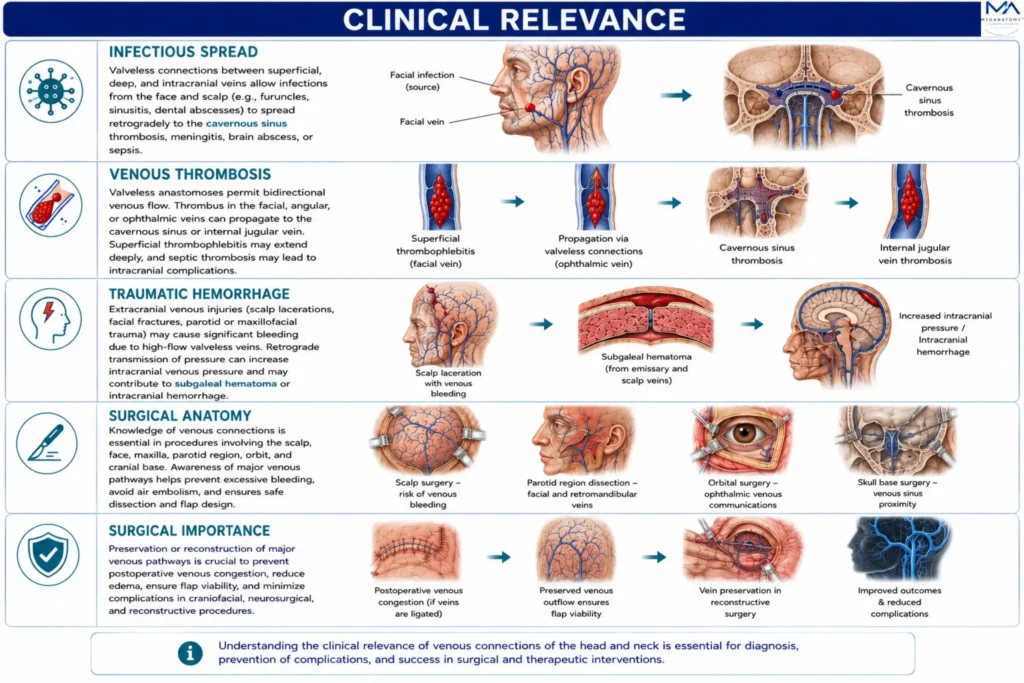
The superficial craniofacial veins constitute the principal extracranial pathways for the propagation of infection because their valveless architecture permits bidirectional venous flow between superficial, deep, and intracranial venous compartments. 

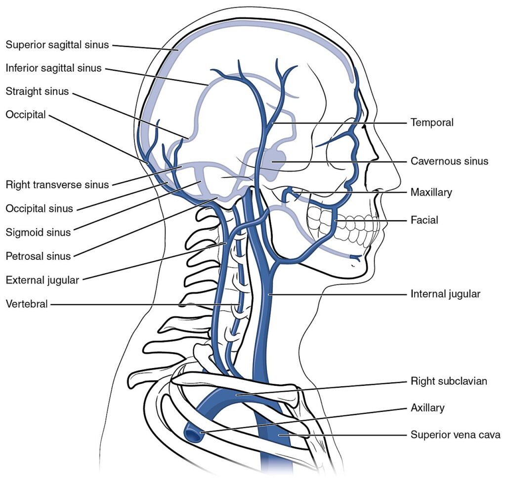
Communications between thefacial, angular, and deep facial veins with the superior and inferior ophthalmic veins, pterygoid venous plexus, and emissary veins allow odontogenic, sinonasal, orbital, and facial soft-tissue infections to extend into the cavernous sinus and other dural venous sinuses. 

Advanced complications include septic cavernous sinus thrombosis, intracranial abscess formation, meningitis, and diffuse intracranial sepsis.

Exam Question

Explain the anatomical pathways responsible for intracranial extension of facial and odontogenic infections through the superficial craniofacial venous system.

Venous Trombosis

The extensive anastomotic organization of the superficial veins permits retrograde propagation of thrombus between superficial and deep venous territories. 

Septic thrombophlebitis involving the facial vein, angular vein, or ophthalmic veins may extend to the cavernous sinus, whereas communications through the deep facial vein and pterygoid venous plexus provide additional routes for thrombotic dissemination. 

Recognition of these anatomical pathways is fundamental for diagnosing cerebral venous sinus thrombosis, differentiating physiological venous enhancement from pathological thrombosis, and planning appropriate medical or endovascular treatment.

Exam Question

How do the anatomical communications of the superficial craniofacial veins influence the development, extension, and diagnosis of venous thrombosis?

Traumatic Hemorrhage

Because the superficial craniofacial veins course within the subcutaneous tissue and superficial musculoaponeurotic system (SMAS), they are highly susceptible to penetrating injury, blunt trauma, facial fractures, and operative dissection. 

Disruption of major venous trunks may produce persistent hemorrhage, hematoma formation, postoperative venous congestion, and compromised soft-tissue perfusion. 

Preservation of dominant venous drainage pathways is therefore essential during craniofacial reconstruction to minimize tissue edema, venous insufficiency, and flap-related complications.

Exam Question

Why are the superficial craniofacial veins particularly vulnerable during facial trauma, and what anatomical factors determine the clinical consequences of venous injury?

Surgical Anatomy

The superficial venous system provides essential anatomical landmarks during parotid, craniofacial, maxillofacial, plastic, microvascular, and reconstructive surgery. 

The retromandibular vein is a key landmark for identifying the facial nerve within the parotid gland, whereas the superficial temporal vein assists orientation during temporal and lateral cranial approaches. 

Accurate preservation or reconstruction of major superficial venous channels optimizes venous drainage, reduces postoperative congestion, and improves tissue viability following free-flap transfer and complex facial reconstruction.

Exam Question

Discuss the surgical importance of the superficial craniofacial veins and explain how their anatomical relationships influence operative planning and reconstructive outcomes.

SUMMARY TABLE

Scroll to Top