Superficial Veins

MYO CORE

Facial Veins

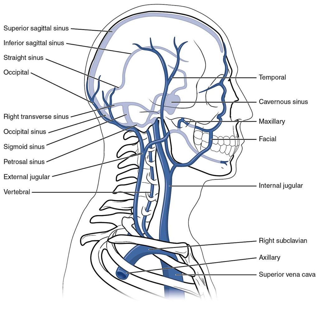
The facial veins form a highly interconnected, valveless superficial venous network that drains the face and establishes extensive communications with the ophthalmic veins, deep facial vein, pterygoid venous plexus, and cervical venous system. These connections create a continuous extracranial–intracranial venous network that supports collateral venous drainage while providing a potential pathway for the spread of infection and septic thrombosis.

OVERVIEW

The facial venous system comprises an extensive superficial valveless plexiform network that drains the superficial tissues of the face and converges into the facial vein, the principal superficial venous channel of the face. 

The facial vein integrates with the angular vein, ophthalmic veins, deep facial vein, pterygoid venous plexus, superficial temporal vein, retromandibular vein, and common facial vein, establishing continuous venous continuity between the facial, cranial, and cervical regions. 

Superiorly, the angular vein communicates with the superior and inferior ophthalmic veins, providing direct continuity with the cavernous sinus, whereas posteriorly the deep facial vein connects the facial vein to the pterygoid venous plexus, linking superficial and deep facial drainage. 

This highly interconnected valveless angioarchitecture forms the structural basis for collateral venous drainage while creating clinically significant extracranial-intracranial pathways for the spread of infection and septic thrombosis.

ANATOMY

Structural Organization

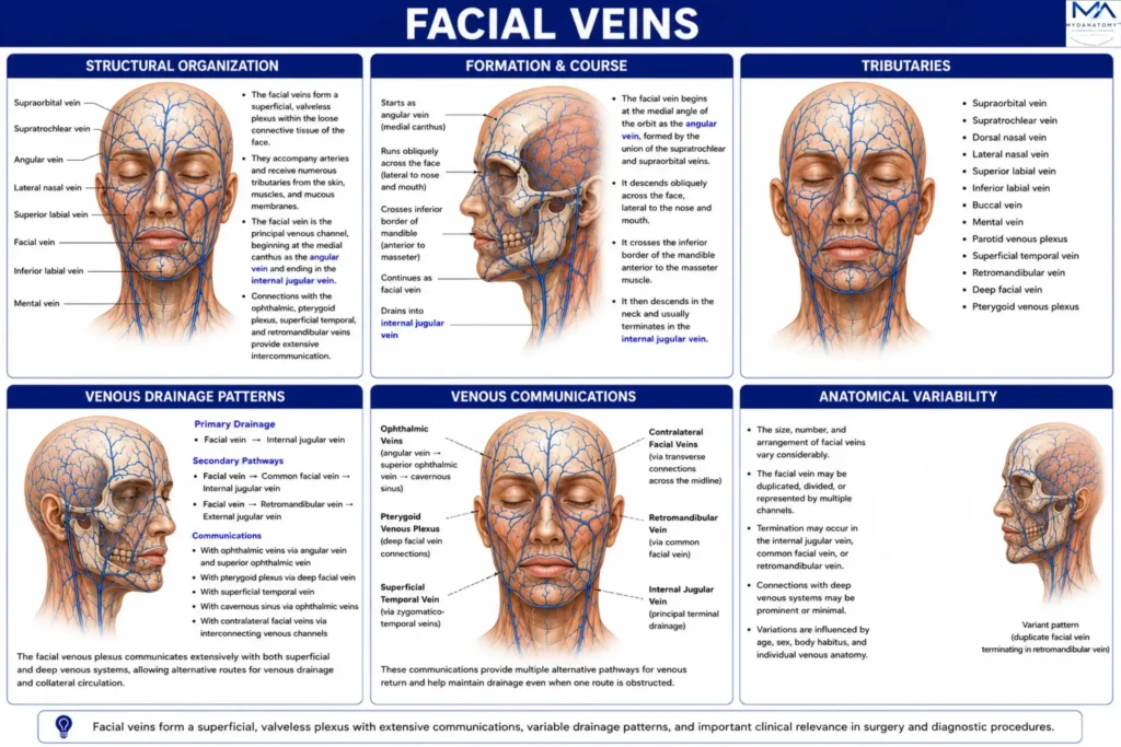
The facial veins are organized as an extensive superficial valveless venous network situated predominantly within the superficial fascia of the face. 

The facial vein constitutes the principal superficial venous channel, receiving numerous cutaneous and muscular tributaries while remaining extensively interconnected with adjacent superficial and deep venous systems. 

Unlike the comparatively constant facial arterial pattern, the facial venous network demonstrates considerable anatomical variability while maintaining continuous collateral communications across the forehead, orbit, nose, cheeks, lips, and chin.

Exam Question

How does the structural organization of the facial venous system integrate the superficial regions of the face into a continuous collateral drainage network, and in what way does it differ from the facial arterial pattern?

Formation & Course

The facial vein begins at the medial angle of the orbit as the continuation of the angular vein, formed predominantly by the union of the supratrochlear and supraorbital veins

It descends obliquely across the face, passing posterior to the facial artery while remaining superficial to the muscles of facial expression and deep to the superficial musculoaponeurotic system (SMAS). 

After crossing the inferior border of the mandible anterior to the masseter muscle, it joins the anterior division of the retromandibular vein to form the common facial vein, which usually terminates in the internal jugular vein.

Exam Question

Describe the formation, anatomical course, topographical relationships, and termination of the facial vein from the angular vein to the internal jugular venous system.

Tributaries

The facial vein receives numerous superficial tributaries corresponding to the arterial distribution of the face. 

Major tributaries include the superior and inferior labial veins, lateral nasal vein, deep facial vein, submental vein, tonsillar vein, and inferior palpebral veins, together with multiple muscular and cutaneous veins. 

These tributaries drain the lips, nose, eyelids, cheeks, chin, muscles of facial expression, and adjacent superficial tissues before converging into the facial vein.

Exam Question

Which major tributaries contribute to the facial vein, and which anatomical regions are drained by each of these venous channels?

Venous Drainage Patterns

Superficial facial venous drainage follows a hierarchical organization in which small cutaneous venules converge into regional tributaries before entering the facial vein. 

The facial vein subsequently drains through the common facial vein into the internal jugular vein, forming the principal superficial venous outflow of the face. 

Additional drainage occurs through communications with the superficial temporal, retromandibular, and external jugular venous systems, providing alternative superficial venous pathways.

Exam Question

Describe the hierarchical organization of superficial facial venous drainage from the cutaneous tributaries to the terminal cervical venous system.

Venous Communications

The facial venous system demonstrates extensive communications with both superficial and deep craniofacial veins. 

Superiorly, the angular vein communicates with the superior and inferior ophthalmic veins, establishing direct continuity with the cavernous sinus

Posteriorly, the deep facial vein connects the facial vein with the pterygoid venous plexus, while lateral communications occur through the superficial temporal and retromandibular veins. 

These extensive valveless communications integrate the superficial face with the deep facial spaces and intracranial venous circulation.

Exam Question

How do the ophthalmic veins, deep facial vein, pterygoid venous plexus, and superficial temporal venous system integrate the superficial facial veins with the deep facial and intracranial venous circulation?

Anatomical Variability

The facial venous system exhibits considerable variation in the caliber, course, termination, and interconnections of its principal veins. 

Variations commonly involve the formation of the common facial vein, communications with the external jugular system, and the development of the angular and deep facial veins. 

Despite these variations, an extensive network of superficial and deep anastomoses consistently preserves venous continuity throughout the face, providing a highly redundant collateral drainage system.

Exam Question

What are the principal anatomical variations of the facial venous system, and how does its extensive anastomotic architecture preserve collateral venous continuity despite variations in venous anatomy?

SUMMARY TABLE

Scroll to Top