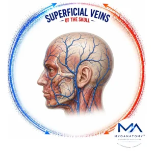
Superficial Veins
MYO CORE
Temporal Veins
The temporal veins form a superficial valveless venous network centered on the superficial temporal vein, integrating venous drainage from the temporal scalp, lateral forehead, auricle, and adjacent facial regions. Through extensive communications with the middle and deep temporal veins, facial vein, maxillary vein, retromandibular vein, and scalp venous plexuses, they establish the principal superficial–deep collateral pathway of the lateral craniofacial venous system.
OVERVIEW
The temporal venous system consists of coordinated superficial and deep venous compartments linked by multiple valveless communications.
The superficial temporal vein receives numerous superficial tributaries and, within the parotid gland, unites with the maxillary vein to form the retromandibular vein, representing the principal transition from superficial cranial to cervical venous drainage.
The middle temporal vein connects the superficial temporal vein with the temporalis region, whereas the deep temporal veins accompany the deep temporal arteries and terminate within the pterygoid venous plexus, establishing the principal anatomical communication between the superficial temporal and deep facial venous systems.
Extensive anastomoses with adjacent scalp and facial veins integrate the temporal region into the continuous superficial venous network of the head.

ANATOMY
Structural Organization
The temporal venous system is organized into superficial and deep valveless venous compartments interconnected by multiple communicating vessels.
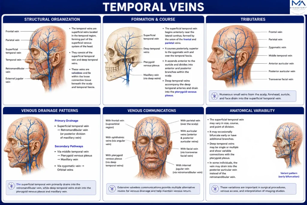
The superficial temporal vein forms the principal superficial venous trunk, whereas the middle temporal vein, deep temporal veins, and pterygoid venous plexus constitute the deep component. These vessels are arranged in a hierarchical architecture that integrates the scalp, temporal region, temporalis muscle, infratemporal fossa, and cervical venous system into a continuous regional network.
This structural organization provides the anatomical framework for coordinated superficial and deep craniofacial venous drainage.
Exam Question
How is the temporal venous system structurally organized, and which anatomical components establish continuity between its superficial and deep venous compartments?
Formation & Course
The superficial temporal vein is formed by the union of the frontal and parietal veins within the temporal scalp.
It descends within the superficial fascia, accompanying the superficial temporal artery anterior to the auricle before crossing the posterior root of the zygomatic arch. The vein then enters the parotid gland, where it joins the maxillary vein to form the retromandibular vein.
Throughout its course, it lies in close relationship to the auriculotemporal nerve, while the middle and deep temporal veins course beneath the temporal fascia on the surface of the temporalis muscle.
Exam Question
Describe the formation, anatomical course, relationships, and termination of the superficial temporal vein.
Tributaries
The superficial temporal vein receives the frontal, parietal, transverse facial, anterior auricular, and middle temporal veins, together with numerous smaller tributaries from the temporal scalp and lateral face.
The middle temporal vein drains the temporalis muscle and temporal fascia before joining the superficial temporal vein, whereas the deep temporal veins accompany the deep temporal arteries and terminate within the pterygoid venous plexus.
Collectively, these tributaries integrate superficial and deep drainage pathways throughout the temporal and infratemporal regions.
Exam Question
Identify the principal tributaries of the temporal venous system and explain how the deep temporal veins integrate superficial and deep venous drainage.
Venous Drainage
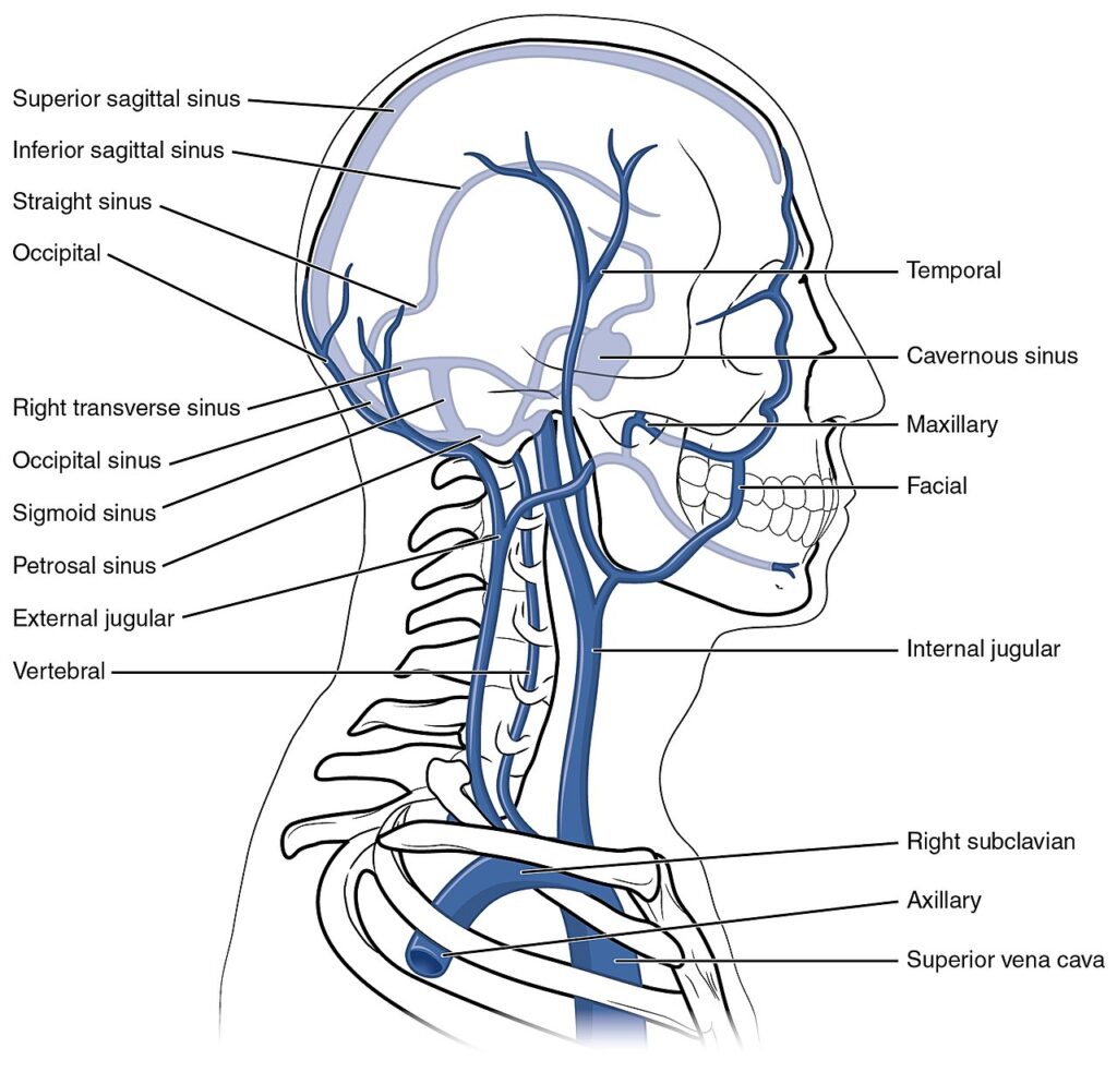
Venous return from the temporal region begins within the superficial tributaries, which converge to form the superficial temporal vein. Within the parotid gland, this vessel joins the maxillary vein to form the retromandibular vein.
The anterior division of the retromandibular vein combines with the facial vein to form the common facial vein, which drains into the internal jugular vein, whereas the posterior division joins the posterior auricular vein to form the external jugular vein.
This arrangement establishes dual pathways for cervical venous drainage.
Exam Question
Trace the complete venous drainage pathway from the temporal region to the internal and external jugular venous systems.
Venous Communications
The temporal venous system forms extensive valveless anastomoses with the frontal, parietal, occipital, posterior auricular, facial, and transverse facial veins, while deeper communications connect with the middle temporal veins, deep temporal veins, and pterygoid venous plexus.
These interconnections establish a continuous collateral network linking the scalp, face, temporal region, and infratemporal fossa.
The resulting venous continuity enables extensive anatomical communication between superficial and deep craniofacial venous systems.
Exam Question
Which superficial and deep venous communications establish collateral continuity throughout the temporal and adjacent craniofacial regions?
Anatomical Variations
The temporal venous system demonstrates considerable variation in the formation, course, caliber, tributary pattern, duplication, termination, and anastomotic configuration of its major veins.
Variability most frequently involves the formation of the superficial temporal vein, the contribution of the middle temporal vein, and the configuration of the retromandibular vein.
Although these variations rarely alter the overall drainage pattern, they are important considerations during temporal, parotid, reconstructive, and endovascular procedures and for accurate interpretation of vascular imaging.

Exam Question
What are the principal anatomical variations of the temporal venous system, and why must they be recognized during surgical and radiological evaluation?
SUMMARY TABLE