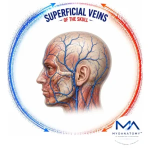
Superficial Veins
MYO CORE
Venous Connections
The venous connections of the head and neck comprise an extensive valveless anastomotic network linking the superficial, deep, extracranial, and intracranial venous systems. These communications establish continuous anatomical continuity between the scalp, face, temporal region, orbit, cranial cavity, and cervical venous systems, forming the principal collateral framework of craniofacial venous organization.
OVERVIEW
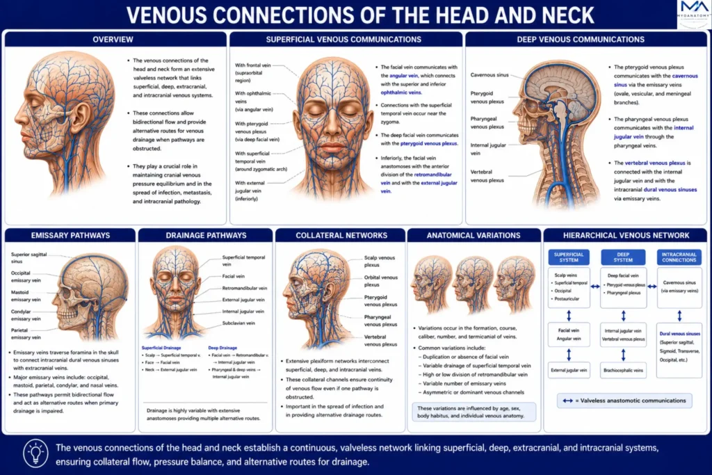
Superficial venous communications interconnect the facial, angular, superficial temporal, occipital, posterior auricular, and external jugular veins, whereas deep communications unite the deep facial vein, ophthalmic veins, pterygoid venous plexus, pharyngeal venous plexus, vertebral venous plexuses, and dural venous sinuses into a continuous craniofacial network.
Emissary and diploic veins establish transosseous communications between the extracranial venous system and the intracranial dural venous sinuses by traversing the cranial foramina and diploë of the skull.
Collectively, these interconnected superficial, deep, osseous, and intracranial pathways constitute a hierarchical angioarchitecture that integrates the principal venous territories of the head and neck while preserving extensive collateral communications.

ANATOMY
Structural Organization
The craniofacial venous connections are organized into hierarchically arranged superficial, deep, extracranial, and intracranial venous compartments united by an extensive network of valveless anastomoses.
Superficial veins, deep venous plexuses, emissary veins, diploic veins, and the dural venous sinuses collectively form a continuous three-dimensional angioarchitecture rather than discrete vascular channels.
This integrated organization establishes uninterrupted anatomical continuity between the scalp, face, orbit, temporal region, cranial cavity, skull, and cervical venous systems, creating the structural framework of craniofacial venous communication.
Exam Question
How is the craniofacial venous communication network structurally organized, and which anatomical components establish continuity between its superficial, deep, extracranial, and intracranial compartments?
Venous Architecture
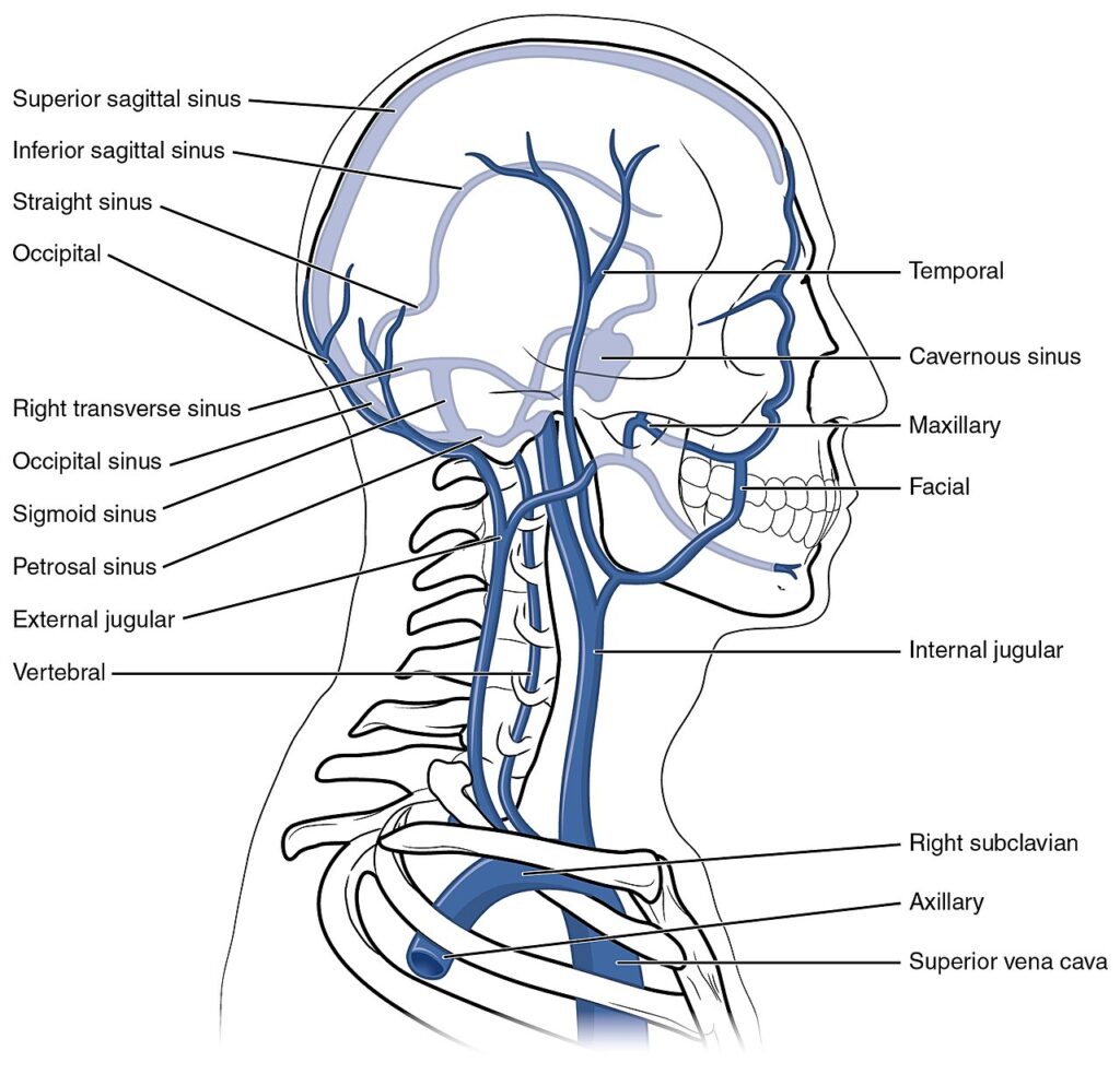
The superficial venous compartment is composed primarily of the facial, superficial temporal, occipital, posterior auricular, and external jugular veins, which communicate extensively through multiple superficial anastomoses.
The deep compartment comprises the deep facial vein, ophthalmic veins, pterygoid venous plexus, pharyngeal venous plexus, vertebral venous plexuses, and dural venous sinuses, interconnected through a dense network of communicating channels.
Together, these superficial and deep components establish a continuous craniofacial venous framework that links extracranial and intracranial venous territories.
Exam Question
Which superficial and deep venous structures constitute the principal anatomical framework of craniofacial venous communications?
Emissary Pathways
Emissary veins are valveless transosseous communicating vessels that traverse the cranial foramina to establish direct anatomical continuity between extracranial veins and the dural venous sinuses.
The principal emissary pathways include the parietal, mastoid, posterior condylar, and occipital emissary veins, each connecting specific extracranial venous territories with the superior sagittal, sigmoid, transverse, or occipital sinuses.
Together with the diploic veins, they form the principal osseous communications between the scalp, skull, and intracranial venous compartments.
Exam Question
Describe the anatomical organization of the emissary veins and explain how they establish transosseous communications between extracranial veins and the dural venous sinuses.
Drainage Pathways
Venous return from the craniofacial region follows coordinated superficial, deep, and intracranial drainage pathways.
Superficial veins converge into the facial, retromandibular, external jugular, and anterior jugular veins, whereas deep venous return is collected by the deep facial vein, pterygoid venous plexus, pharyngeal venous plexus, and vertebral venous plexuses, ultimately draining into the internal jugular or vertebral venous systems.
Intracranially, cerebral and meningeal veins empty into the dural venous sinuses, which terminate primarily at the internal jugular veins through the jugular foramina.
Exam Question
Trace the principal superficial, deep, and intracranial venous drainage pathways of the craniofacial venous communication network.
Collateral Networks
The craniofacial venous system is characterized by an extensive collateral network formed through valveless anastomoses between the facial, angular, ophthalmic, deep facial, superficial temporal, occipital, posterior auricular, and vertebral venous systems.
Additional communications through the pterygoid venous plexus, emissary veins, and diploic veins integrate superficial, deep, osseous, and intracranial venous compartments.
These collateral pathways establish uninterrupted anatomical continuity throughout the scalp, skull, face, orbit, cranial cavity, and cervical region.

Exam Question
Which principal collateral pathways establish anatomical continuity between the superficial, deep, osseous, extracranial, and intracranial craniofacial venous systems?
Anatomical Variations
The craniofacial venous communications demonstrate considerable variation in number, caliber, course, termination, anastomotic configuration, and regional dominance.
Variability most frequently involves the facial–ophthalmic, deep facial–pterygoid, superficial temporal, vertebral, and emissary venous pathways, as well as the development of individual diploic and communicating veins.
Although the overall organizational pattern remains conserved, recognition of these variations is essential for craniofacial, neurosurgical, maxillofacial, otologic, and reconstructive procedures and for accurate interpretation of angiographic and cross-sectional imaging.
Exam Question
What are the principal anatomical variations of the craniofacial venous communications, and how do they influence surgical planning and radiological interpretation?
SUMMARY TABLE