Superficial Veins

MYO CORE

Scalp Veins

The scalp veins form a highly interconnected, valveless superficial venous network that communicates with the intracranial dural venous sinuses through emissary veins. Their extensive anastomoses maintain collateral drainage while providing a potential route for the spread of infection or thrombosis. Venous return ultimately drains into the internal and external jugular venous systems.

OVERVIEW

The scalp venous system comprises a dense, valveless superficial plexiform network embedded within the connective tissue layer of the scalp, where fibrous septa firmly anchor the veins alongside the scalp arteries. 

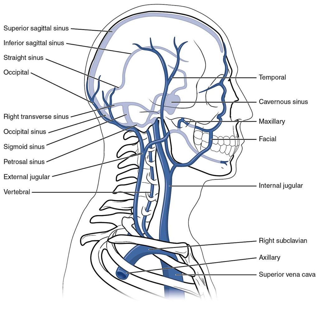
Extensive longitudinal and transverse anastomoses integrate the frontal, temporal, parietal, and occipital regions into a continuous collateral network that converges with the facial, retromandibular, external jugular, and internal jugular venous systems. 

Valveless emissary veins establish direct communications with the intracranial dural venous sinuses, creating a continuous extracranial–intracranial venous continuum that permits bidirectional flow in response to physiological pressure gradients.

This specialized venous architecture optimizes collateral venous return and pressure redistribution while providing a clinically significant conduit for the intracranial spread of infection, thrombosis, and inflammatory processes.

ANATOMY

Structural Organization

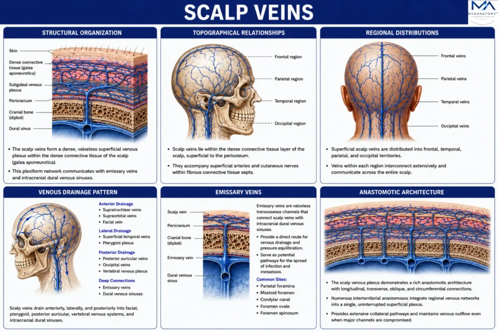
The scalp veins are organized as a superficial valveless plexiform venous network situated predominantly within the dense connective tissue layer of the scalp. 

Numerous longitudinal, transverse, oblique, and cross-midline venous channels interconnect to form a continuous superficial angioarchitecture rather than discrete segmental vessels. 

Progressive convergence of smaller tributaries forms larger regional collecting veins while preserving uninterrupted collateral continuity between the frontal, temporal, parietal, and occipital regions. This multidirectional plexiform arrangement constitutes the principal structural framework of superficial scalp venous drainage.

Exam Question

How does the multidirectional plexiform architecture of the superficial scalp veins establish continuous collateral drainage while preserving anatomical integration between all scalp regions?

Topographical Relationships

The scalp veins lie within the dense connective tissue (second) layer of the scalp, where they accompany the corresponding arteries and cutaneous nerves within fibrous connective tissue septa extending from the dermis to the epicranial aponeurosis. 

Firm attachment of the venous walls to the surrounding connective tissue limits vascular displacement after injury and maintains stable neurovascular relationships throughout the scalp. 

Their superficial position and close association with adjacent arteries define the characteristic topographical organization of the scalp neurovascular bundles.

Exam Question

How do the topographical relationships of the scalp veins within the dense connective tissue layer determine their anatomical fixation, neurovascular organization, and surgical significance?

Regional Distributions

The superficial scalp veins are distributed into frontal, temporal, parietal, and occipital venous territories, each drained by dominant regional veins while remaining extensively interconnected through superficial venous communications. 

The frontal scalp drains through the supratrochlear and supraorbital veins, the temporal region through the superficial temporal vein, and the posterior scalp through the posterior auricular and occipital veins. 

Despite regional predominance, abundant interterritorial anastomoses eliminate distinct venous boundaries and integrate the entire scalp into a continuous superficial drainage network.

Exam Question

How are the regional venous territories of the scalp anatomically organized, and how do their interterritorial communications integrate the scalp into a continuous superficial venous network?

Venous Drainage Pattern

Venous drainage follows a hierarchical anatomical pattern, beginning with superficial cutaneous tributaries that converge into regional collecting veins before terminating within the major cervical venous channels. 

The supratrochlear and supraorbital veins drain anteriorly into the angular and facial veins, whereas the superficial temporal vein joins the maxillary vein to form the retromandibular vein. 

Posterior drainage proceeds through the posterior auricular and occipital veins before ultimately emptying into the external and internal jugular venous systems, completing superficial cranial venous outflow.

Exam Question

Describe the hierarchical anatomical organization of superficial scalp venous drainage from the primary cutaneous tributaries to its terminal cervical venous pathways.

Emissary Veins

The emissary veins are valveless transosseous vessels that traverse cranial foramina to establish direct anatomical communications between the superficial scalp veins and the intracranial dural venous sinuses. 

The principal emissary pathways include the parietal, mastoid, occipital, and condylar emissary veins, which communicate predominantly with the superior sagittal, transverse, and sigmoid sinuses. 

These vessels constitute the principal extracranial–intracranial venous connections and represent an integral component of cranial venous anatomy.

Exam Question

How do the emissary veins establish anatomical continuity between the superficial scalp veins and the intracranial dural venous sinuses, and which emissary pathways provide the principal extracranial–intracranial communications?

Anastomotic Architecture

The scalp veins demonstrate an exceptionally rich superficial anastomotic architecture composed of longitudinal, transverse, oblique, circumferential, and cross-midline venous communications. These multidirectional anastomoses integrate all regional drainage territories into a single uninterrupted superficial venous plexus extending across the entire scalp. 

This highly redundant organization provides the anatomical basis for extensive collateral continuity and distinguishes the scalp venous system from the more segmental venous arrangements observed in many other anatomical regions.

Exam Question

How does the multidirectional anastomotic architecture of the superficial scalp veins maintain uninterrupted collateral continuity, and in what ways does it differ from segmental venous organization elsewhere in the body?

SUMMARY TABLE

Scroll to Top