Sacral Plexus
MYO CORE
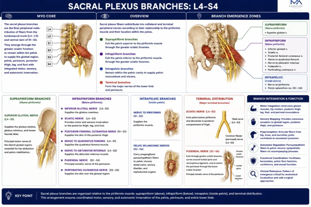
Branches
The sacral plexus branches comprise the collateral and terminal peripheral nerves that emerge from the plexus in a highly organized topographical pattern. Classified according to their relationship to the piriformis muscle and the pelvic cavity, they provide integrated motor, sensory, proprioceptive, and autonomic innervation to the gluteal region, pelvis, perineum, and nearly the entire lower limb.
OVERVIEW
The sacral plexus distributes its fibers into collateral and terminal peripheral branches according to their anatomical relationship to the piriformis muscle and their course within or beyond the pelvic cavity.
The suprapiriform branch, represented by the superior gluteal nerve, exits the pelvis superior to the piriformis through the greater sciatic foramen.
The infrapiriform branches –including the inferior gluteal, sciatic, posterior femoral cutaneous, nerve to quadratus femoris, nerve to obturator internus, pudendal, and perforating cutaneous nerves-emerge inferior to the piriformis through the greater sciatic foramen to supply the gluteal region, posterior thigh, lower limb, and perineum.
Intrapelvic branches, comprising the nerve to piriformis and pelvic splanchnic nerves, remain within the pelvic cavity to innervate pelvic musculature and pelvic viscera. Distally, the terminal branches, principally the sciatic and pudendal nerves, establish the major neural pathways of the lower limb and perineum.
This highly ordered branching architecture generates functionally specialized peripheral nerves that integrate motor, sensory, proprioceptive, and autonomic functions, ensuring coordinated regional innervation while providing essential anatomical landmarks for neurological localization, regional anesthesia, and pelvic and gluteal surgical approaches.
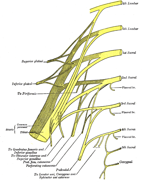
Plan of sacral and pudendal plexuses (Gray828)” – Henry Vandyke Carter, Gray’s Anatomy: Anatomy of the Human Body. Source: Wikimedia Commons. Public Domain.
Exam Question
Describe the anatomical organization of the sacral plexus branches according to their relationship to the piriformis muscle and the pelvic cavity. Differentiate the suprapiriform, infrapiriform, intrapelvic, and terminal branches, and explain how their topographical arrangement determines regional motor, sensory, proprioceptive, and autonomic innervation as well as their importance in neurological localization, regional anesthesia, and pelvic and gluteal surgery.
ANATOMY
Branch Classification
The branches of the sacral plexus are anatomically classified according to their topographical relationship to the piriformis muscle and their course within the pelvic cavity into suprapiriform, infrapiriform, intrapelvic, and terminal branches.
This classification reflects the orderly peripheral redistribution of multisegmental nerve fibers into distinct neuroanatomical pathways supplying the pelvis and lower limb.
Their predictable arrangement establishes the anatomical basis for regional motor, sensory, proprioceptive, and autonomic innervation while providing reliable landmarks for neurological localization, regional anesthesia, and pelvic surgery.
Exam Question
Describe the anatomical classification of the sacral plexus branches according to their relationship to the piriformis muscle and the pelvic cavity, and explain how this topographical organization determines regional innervation and its clinical significance.
Suprapiriform Branches
The superior gluteal nerve (L4- S1) is the sole suprapiriform branch, emerging superior to the piriformis through the greater sciatic foramen.
It supplies the gluteus medius, gluteus minimus, and tensor fasciae latae, constituting the principal motor nerve of the lateral gluteal compartment.
By coordinating hip abduction, medial rotation, and dynamic pelvic stabilization during the stance phase of gait, it maintains frontal-plane pelvic alignment and efficient locomotor biomechanics.
Exam Question
Explain the anatomical course, spinal root contributions, muscular distribution, and biomechanical importance of the superior gluteal nerve as the sole suprapiriform branch of the sacral plexus.
Infrapiriform Branches
The infrapiriform compartment contains the inferior gluteal nerve (L5–S2), sciatic nerve (L4–S3), posterior femoral cutaneous nerve (S1–S3), nerve to quadratus femoris (L4–S1), nerve to obturator internus (L5–S2), pudendal nerve (S2–S4), and perforating cutaneous nerve (S2–S3), all of which emerge inferior to the piriformis through the greater sciatic foramen.
Collectively, these branches establish the principal somatic neural pathways of the gluteal region, posterior thigh, and lower limb, while providing somatic and autonomic continuity to the perineum through the pudendal nerve and associated pelvic autonomic pathways.
Exam Question
Identify the infrapiriform branches of the sacral plexus and discuss their anatomical course, regional distribution, and collective functional significance in the innervation of the gluteal region, lower limb, and perineum.
Intrapelvic Branches
The intrapelvic branches remain entirely within the pelvic cavity and include the nerve to piriformis (S1–S2) and the pelvic splanchnic nerves (S2–S4).
The nerve to piriformis provides the exclusive motor supply to the piriformis muscle, whereas the pelvic splanchnic nerves constitute the sacral parasympathetic outflow, conveying preganglionic parasympathetic fibers through the inferior hypogastric plexus to the distal colon, rectum, urinary bladder, pelvic urethra, erectile tissues, and reproductive organs.
These fibers regulate visceral smooth muscle activity, glandular secretion, micturition, defecation, and sexual function.
Exam Question
Describe the intrapelvic branches of the sacral plexus, emphasizing their anatomical course, fiber composition, and physiological roles in somatic and pelvic autonomic function.
Terminal Branches
The sciatic nerve (L4–S3) and pudendal nerve (S2–S4) represent the principal terminal continuations of the sacral plexus.
The sciatic nerve exits inferior to the piriformis through the greater sciatic foramen, descends through the posterior thigh, and typically divides within the distal thigh or at the apex of the popliteal fossa into the tibial and common fibular (peroneal) nerves, although the level of division is anatomically variable.
The pudendal nerve exits through the greater sciatic foramen, passes around the ischial spine and sacrospinous ligament, and re-enters the perineum through the lesser sciatic foramen before traversing the pudendal canal.
Together, these terminal branches establish the principal somatic neural supply of the lower limb and perineum.
Exam Question
Discuss the anatomical course, branching pattern, and functional distribution of the sciatic and pudendal nerves as the principal terminal branches of the sacral plexus.
Branching Integration
The branching pattern of the sacral plexus integrates collateral and terminal nerves into a coordinated peripheral network supplying the pelvis, perineum, and lower limb.
Motor branches coordinate movement of the gluteal region, hip, posterior thigh, leg, foot, pelvic floor, and perineum, whereas sensory and proprioceptive fibers provide cutaneous sensation and joint position feedback essential for posture and locomotion.
Autonomic branches regulate pelvic visceral function through parasympathetic and sympathetic pathways.
This integrated branching architecture ensures coordinated somatic and visceral function while providing a precise anatomical framework for neurological localization, regional anesthesia, and pelvic, gluteal, and lower-limb surgery.
Exam Question
Explain how the branching pattern of the sacral plexus integrates motor, sensory, proprioceptive, and autonomic pathways to coordinate pelvic and lower-limb function, and discuss its anatomical and clinical significance.
SUMMARY TABLE