Sacral Plexus
MYO CORE
Clinical Significance
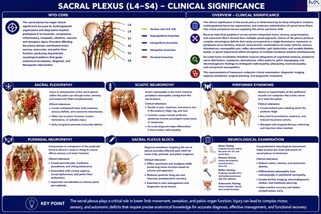
The sacral plexus has major clinical significance because its multisegmental organization and deep pelvic location predispose it to traumatic, compressive, inflammatory, neoplastic, obstetric, vascular, and iatrogenic injury. Disorders affecting the plexus disrupt coordinated motor, sensory, autonomic, and pelvic floor function, producing characteristic neurological patterns that guide anatomical localization, diagnosis, and therapeutic intervention.
OVERVIEW
The clinical significance of the sacral plexus is determined by its deep intrapelvic location, multisegmental fascicular organization, and extensive redistribution of spinal nerve fibers into mixed peripheral nerves supplying the pelvis and lower limb.
Because individual peripheral nerves contain integrated motor, sensory, proprioceptive, and autonomic fibers derived from multiple spinal segments, lesions of the plexus produce complex neurological deficits that rarely correspond to a single dermatome, myotome, or peripheral nerve territory. Instead, characteristic combinations of motor deficits, sensory disturbances, neuropathic pain, reflex abnormalities, gait dysfunction, and variable bladder, bowel, or sexual impairment reflect disruption of multiple functional systems simultaneously.
Accurate lesion localization therefore requires integration of segmental anatomy, peripheral nerve distribution, myotomes, dermatomes, reflex patterns, pelvic topography, and electrodiagnostic findings to distinguish radiculopathy, plexopathy, mononeuropathy, and entrapment neuropathies.
This neuroanatomical framework underpins clinical examination, diagnostic imaging, regional anesthesia, surgical planning, and prognostic evaluation.
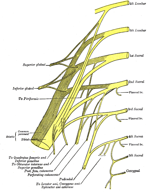
Plan of sacral and pudendal plexuses (Gray828)” – Henry Vandyke Carter, Gray’s Anatomy: Anatomy of the Human Body. Source: Wikimedia Commons. Public Domain.
Exam Question
Explain how the multisegmental fascicular organization of the sacral plexus determines the neurological manifestations of plexopathy and peripheral nerve injury, and discuss its importance in neuroanatomical localization, differential diagnosis, and clinical management.
ANATOMY
Sacral Plexopathy
Sacral plexopathy results from injury or pathological involvementof the sacral plexus within the posterior pelvis.
Common causes include pelvic fractures, penetrating trauma, neoplasms, radiation-induced fibrosis, inflammatory plexopathies, compressive hematomas, vascular lesions, obstetric trauma, and iatrogenic injury. Because the plexus integrates fibers from multiple spinal segments into mixed peripheral nerves, lesions produce combined motor, sensory, reflex, and autonomic deficits across multiple peripheral nerve territories.
Patients typically present with lower-limb weakness, sensory impairment, neuropathic pain, diminished reflexes, gait disturbance, and variable bladder, bowel, or sexual dysfunction. This multisegmental neurological pattern distinguishes sacral plexopathy from isolated mononeuropathy and lumbosacral radiculopathy.
Exam Question
Discuss the anatomical basis, etiologies, neurological manifestations, and diagnostic features of sacral plexopathy, and explain how its multisegmental pattern differentiates it from radiculopathy and isolated peripheral nerve injury.
Sciatic Neuropathy
Sciatic neuropathy is the most common major peripheral neuropathy originating from the sacral plexus.
Injury may result from hip dislocation, pelvic fractures, penetrating trauma, gluteal intramuscular injection, prolonged compression, neoplastic infiltration, or iatrogenic injury.
Clinical deficits depend on the level of injury relative to the sciatic bifurcation and include weakness of knee flexion, paralysis of muscles below the knee, sensory loss of the leg and foot, neuropathic pain, diminished ankle reflexes, and characteristic gait abnormalities such as foot drop.
Anatomical localization requires differentiation from lumbosacral radiculopathy, common fibular neuropathy, tibial neuropathy,and distal sciatic lesions.
Exam Question
Explain how the anatomical course and level of injury of the sciatic nerve determine the motor, sensory, and clinical manifestations of sciatic neuropathy, and discuss its differentiation from radiculopathy and distal peripheral nerve lesions.
Piriformis Syndrome
Piriformis syndrome results from compression or irritation of the sciatic nerve as it passes inferior to-or less commonly through-the piriformis muscle within the greater sciatic foramen.
Predisposing factors include muscular hypertrophy, inflammation, fibrosis, repetitive overuse, pelvic trauma, and anatomical variation of the sciatic nerve. Patients typically develop deep gluteal pain radiating along the sciatic distribution, often aggravated by prolonged sitting or hip flexion, adduction, and internal rotation.
Knowledge of the anatomical relationship between the piriformis muscle and sciatic nerve is essential for distinguishing piriformis syndrome from lumbar disc disease, sacral radiculopathy, and other causes of sciatica.
Exam Question
Describe the anatomical relationship between the piriformis muscle and the sciatic nerve, and discuss the pathophysiological basis, clinical presentation, and differential diagnosis of piriformis syndrome.
Pudendal Neuropathy
Pudendal neuropathy most commonly results from compression of the pudendal nerve at the ischial spine, beneath the sacrospinous ligament, or within the pudendal (Alcock’s) canal. Common causes include prolonged cycling, childbirth, pelvic trauma, chronic compression, fibrosis, and pelvic surgery.
Clinical manifestations include chronic perineal pain, sensory disturbance, sphincter dysfunction, sexual dysfunction, and impaired pelvic floor control.
Because the pudendal nerve is the principal somatic nerve of the perineum, detailed knowledge of its anatomical course and entrapment sites is essential for diagnosis, image-guided nerve blockade, surgical decompression, and pain management.
Exam Question
Explain the anatomical course of the pudendal nerve and discuss how its sites of entrapment determine the clinical manifestations, diagnosis, and management of pudendal neuropathy.
Sacral Plexus Block
Sacral plexus block is an advanced regional anesthetic technique that temporarily interrupts neural conduction within the sacral plexus to provide anesthesia and postoperative analgesia for procedures involving the posterior thigh, knee, leg, ankle, and foot.
Successful blockade depends on accurate identification of the greater sciatic foramen, piriformis muscle, ischial spine, and adjacent neurovascular structures.
Ultrasound guidance combined with peripheral nerve stimulation improves needle placement, anesthetic distribution, procedural efficacy, and patient safety while reducing complications such as vascular puncture, intraneural injection, local anesthetic toxicity, and incomplete blockade.
Exam Question
Discuss the anatomical principles underlying sacral plexus block and explain how detailed knowledge of pelvic anatomy improves procedural accuracy, anesthetic efficacy, and patient safety.
Neurological Examination
Neurological assessment of the sacral plexus integrates motor testing, sensory mapping, reflex evaluation, gait analysis, autonomic assessment, and targeted provocative maneuvers with detailed knowledge of myotomes, dermatomes, peripheral nerve territories, and segmental anatomy.
Correlation of these findings enables precise localization of lesions to the lumbosacral roots, sacral plexus, or individual peripheral nerves, facilitating differentiation between radiculopathy, plexopathy, mononeuropathy, and entrapment neuropathies.
This systematic neuroanatomical approach underpins electrodiagnostic evaluation, imaging, therapeutic planning, surgical decision-making, and prognostic assessment.
Exam Question
Explain how neurological examination of the sacral plexus integrates motor, sensory, reflex, autonomic, and segmental findings to localize lesions and differentiate radiculopathy, plexopathy, mononeuropathy, and entrapment neuropathies.
SUMMARY TABLE