Lumbar Plexus
MYO CORE

Formation
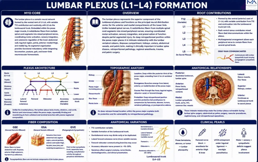
The lumbar plexus is fusion of the ventral rami of L1–L4 spinal nerves, with variable Th12 contribution and continuity with L5 through the lumbosacral trunk. Within the psoas major, it redistributes GSE, GSA, and accompanying postganglionic sympathetic (GVE) fibers to coordinate motor, sensory, and autonomic innervation of the lower abdominal wall and anterior-medial lower limb.
OVERVIEW
The lumbar plexus forms the superior component of the lumbosacral plexus and constitutes the principal somatic neural network of the posterior abdominal wall and anterior-medial lower limb.
Formed by the intercommunication of the ventral rami of L1–L4, it redistributes segmental spinal nerve fibers into mixed peripheral nerves that integrate voluntary motor control, somatic sensation, proprioceptive feedback, and segmental reflex activity.
Embedded within the psoas major muscle, the plexus establishes functional continuity with the sacral plexus through the lumbosacral trunk, ensuring coordinated neuromuscular control of the pelvis and lower extremity.
Its deep retroperitoneal position places it in close relationship to the lumbar vertebral column, iliopsoas compartment, kidneys, ureters, major abdominal vessels, and pelvic brim, making it particularly susceptible to compression, trauma, retroperitoneal pathology, and iatrogenic injury during spinal, vascular, abdominal, and pelvic procedures.
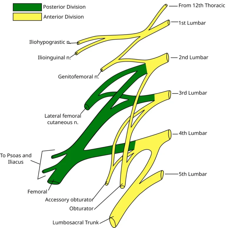
Lumbar Plexus. Adapted from Gray822_es.svg by Gray; derivative work by Ninovolador and Mcstrother. Licensed under CC BY 3.0 via Wikimedia Commons.
Exam Question
Explain how the structural organization, intersegmental fiber redistribution, and retroperitoneal topography of the lumbar plexus establish coordinated neuromuscular control of the pelvis and lower limb while providing the anatomical basis for lesion localization and surgical risk assessment.
ANATOMY
Roots Contribution
The lumbar plexus is formed by the ventral (anterior) rami of L1–L4, with variable contribution from Th12 superiorly and continuation into the lumbosacral trunk (L4–L5) inferiorly.
Within the substance of the psoas major, these ventral rami undergo extensive segmental intercommunication and axonal redistribution before forming peripheral nerves.
Consequently, individual nerves contain fibers derived from multiple spinal cord segments, providing functional overlap, coordinated neuromuscular control, and preservation of motor and sensory function following partial radicular injury.
Exam Question
How do the segmental root contributions and intraplexal redistribution of the lumbar plexus establish functional redundancy while influencing the neurological deficits produced by isolated lumbar radiculopathy?
Plexus Architecture
Unlike the brachial plexus, the lumbar plexus does not organize into distinct trunks, divisions, or cords. Instead, it forms a compact intramuscular network of communicating ventral rami embedded within the posterior part of the psoas major.
Segmental fibers repeatedly converge, diverge, and redistribute before giving rise to collateral and terminal branches, preserving overlapping spinal segment representation while ensuring efficient peripheral nerve formation, coordinated neuromuscular activation, and functional continuity of the anterior abdominal wall, pelvis, and lower limb.
Exam Question
How does the intramuscular segmental architecture of the lumbar plexus optimize peripheral nerve formation, overlapping innervation, and preservation of neurological function following partial plexus injury?
Topographical Anatomy
The lumbar plexus occupies a deep retroperitoneal position within the posterior third of the psoas major muscle, extending approximately from the L1 to L4 vertebral levels.
Its branches emerge from the lateral, anterior, and medial borders of the muscle before traversing the iliac fossa, inguinal region, obturator canal, and femoral triangle to reach their target structures.
Although this deep location provides mechanical protection, it also places the plexus within a confined retrofascial compartment where expanding hematomas, psoas abscesses, retroperitoneal tumors, vertebral pathology, and postoperative collections may produce compressive lumbar plexopathy.
Exam Question
How does the retroperitoneal topography of the lumbar plexus determine both its protected anatomical position and its susceptibility to compressive lumbar plexopathy?
Anatomical Relations
The lumbar plexus is embedded within the psoas major, immediately anterior to the lumbar transverse processes and vertebral bodies.
It is closely related posteriorly to the quadratus lumborum and lumbar spine; medially to the lumbar sympathetic trunk, abdominal aorta, inferior vena cava, and common iliac vessels; laterally to the iliacus and iliac fascia; and superiorly to the kidneys and ureters. Inferiorly, its continuation through the lumbosacral trunk establishes direct anatomical continuity with the sacral plexus.
These intimate relationships explain the vulnerability of the plexus during lumbar spinal procedures, retroperitoneal surgery, vascular reconstruction, nephrectomy, pelvic operations, and traumatic retroperitoneal hemorrhage.
Exam Question
How do the anatomical relationships of the lumbar plexus with adjacent musculoskeletal, vascular, and retroperitoneal structures influence lesion localization, surgical exposure, and operative risk?
Fiber Composition
The lumbar plexus contains general somatic efferent (GSE) fibers supplying the lower abdominal wall, iliopsoas, anterior thigh, and medial thigh musculature; general somatic afferent (GSA) fibers transmitting cutaneous, proprioceptive, and articular sensation from the lower abdomen, inguinal region, external genitalia, anterior-medial thigh, hip, knee, and medial leg; and accompanying postganglionic sympathetic general visceral efferent (GVE) fibers that regulate vasomotor tone, sudomotor activity, and pilomotor function throughout its peripheral distribution.
Parasympathetic fibers are not intrinsic components of the lumbar plexus but reach pelvic viscera through the sacral plexus and pelvic splanchnic nerves.
Exam Question
How do the distributions of GSE, GSA, and accompanying postganglionic sympathetic GVE fibers collectively determine the motor, sensory, and autonomic manifestations of lumbar plexus lesions?
Anatomical Variations
Although the overall organization of the lumbar plexus is highly conserved, considerable variation exists in its segmental contributions, branching patterns, and peripheral communications.
Common variants include contribution from Th12, variable formation of the lumbosacral trunk, duplication or early division of the genitofemoral nerve, variable courses of the lateral femoral cutaneous nerve, femoral–obturator communicating branches, and the presence of an accessory obturator nerve in approximately 10–30% of individuals.
Recognition of these anatomical variations is essential during lumbar plexus blockade, retroperitoneal and pelvic surgery, peripheral nerve reconstruction, and electrodiagnostic interpretation, as atypical branching patterns may substantially alter clinical presentation, surgical anatomy, and procedural outcomes.
Exam Question
How can variations in the formation and branching pattern of the lumbar plexus influence regional anesthesia, surgical planning, electrodiagnostic interpretation, and the clinical presentation of lumbar neuropathies?
SUMMARY TABLE