Lumbar Plexus
MYO CORE

Branches
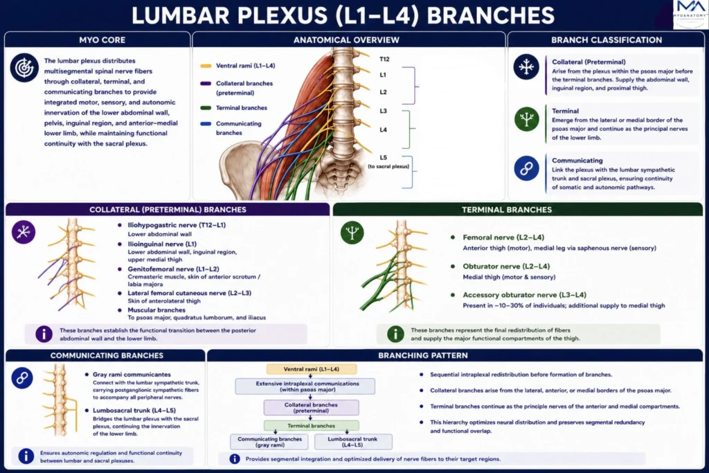
The lumbar plexus distributes multisegmental spinal nerve fibers through collateral, terminal, and communicating branches to establish integrated motor, sensory, and autonomic innervation of the lower abdominal wall, pelvis, inguinal region, and anterior-medial lower limb. Its hierarchical branching pattern ensures coordinated locomotion, postural stability, and functional continuity with the sacral plexus.
OVERVIEW
The lumbar plexus undergoes extensive intraplexal redistribution within the psoas major, transforming segmental spinal nerve roots into an integrated network of collateral, terminal, and communicating branches.
Consequently, individual peripheral nerves contain fibers derived from multiple lumbar spinal segments rather than a single root, establishing overlapping motor and sensory territories that enhance functional integration and neurological resilience.
Through its communications with the lumbar sympathetic trunk and the lumbosacral trunk, the plexus forms a continuous neural interface between the lumbar and sacral regions, coordinating somatic and autonomic innervation of the lower abdominal wall, pelvis, and lower limb.
This highly organized branching architecture provides the anatomical foundation for efficient locomotion, postural stability, proprioceptive control, and coordinated neurovascular regulation while minimizing functional impairment following partial nerve or root injury.
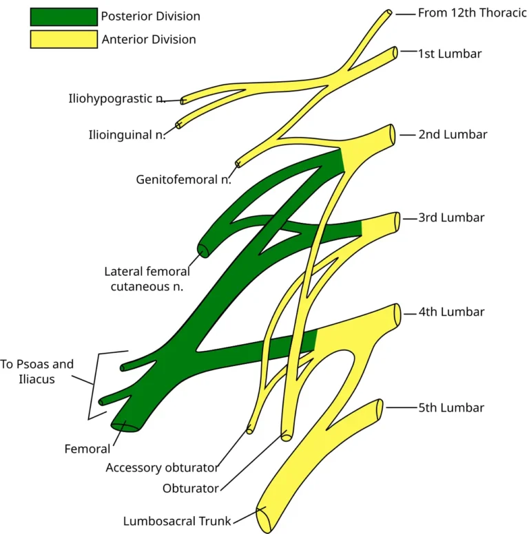
Lumbar Plexus. Adapted from Gray822_es.svg by Gray; derivative work by Ninovolador and Mcstrother. Licensed under CC BY 3.0 via Wikimedia Commons.
Exam Question
How do intraplexal redistribution and communication with the lumbar sympathetic and sacral plexuses establish the functional organization of the lumbar plexus and determine its clinical behavior following neurological injury?
ANATOMY
Branch Classification
The lumbar plexus is organized into collateral, terminal, and communicating branches according to their developmental sequence, topographical origin, and functional distribution.
Following extensive intraplexal communication within the psoas major, fibers from adjacent ventral rami are redistributed into peripheral nerves that emerge from the lateral, anterior, or medial borders of the muscle according to their embryological destination.
This hierarchical branching organization transforms segmental spinal nerve roots into integrated peripheral nerves while preserving multisegmental continuity, functional specialization, and coordinated somatic innervation throughout the lower abdomen, pelvis, and lower limb.
Exam Question
How does the hierarchical classification of lumbar plexus branches reflect intraplexal fiber redistribution, embryological organization, and functional specialization of the lumbosacral nervous system?
Collateral Branches
Collateral branches arise directly from the lumbar plexus before formation of its terminal nerves and emerge predominantly from the lateral and anterior borders of the psoas major.
They include the iliohypogastric (Th12–L1), ilioinguinal (L1), genitofemoral (L1–L2), and lateral femoral cutaneous (L2–L3) nerves, together with segmental muscular branches supplying the psoas major, quadratus lumborum, and iliacus.
These nerves establish the functional transition between the posterior abdominal wall and proximal lower limb by integrating muscular stabilization, abdominal wall competence, inguinal support, cutaneous sensation, and proprioceptive input before the emergence of the major terminal nerves.
Exam Question
How do the anatomical origin, emergence, and functional distribution of the collateral branches establish the transition between the posterior abdominal wall, pelvis, and proximal lower limb?
Terminal Branches
The femoral nerve arises predominantly from the posterior divisions of the ventral rami of L2–L4, whereas the obturator nerve originates from the anterior divisions of L2–L4. The accessory obturator nerve (L3–L4), when present, represents an additional anterior division derivative.
These terminal branches constitute the definitive peripheral continuation of the lumbar plexus, integrating multisegmental fibers into the principal nerves supplying the anterior and medial compartments of the thigh.
Beyond muscular innervation, they convey articular branches to the hip and knee, proprioceptive afferents essential for locomotor control, and overlapping sensory territories that preserve neurological function following partial root or plexus injury.
Exam Question
How do the anterior and posterior divisions of the lumbar plexus determine the anatomical formation, functional distribution, and clinical deficits associated with the femoral and obturator nerves?
Communicating Branches
The lumbar plexus communicates medially with the lumbar sympathetic trunk through gray rami communicantes, allowing postganglionic sympathetic fibers to accompany every peripheral branch and regulate vasomotor tone, sudomotor activity, and pilomotor responses.
Inferiorly, the descending portion of the L4 ventral ramus joins the L5 ventral ramus to form the lumbosacral trunk, the principal anatomical bridge between the lumbar and sacral plexuses.
These communications establish uninterrupted integration between somatic and autonomic pathways while coordinating neural transmission across the pelvis and lower extremity.
Exam Question
How do the gray rami communicantes and lumbosacral trunk integrate somatic and sympathetic pathways to establish functional continuity throughout the lumbosacral plexus?
Regional Distribution
The branches of the lumbar plexus collectively supply the posterior abdominal wall, iliopsoas compartment, inguinal region, external genitalia, pelvic wall, hip joint, anterior and medial thigh, knee joint, and medial leg through a highly organized pattern of overlapping peripheral innervation.
This distribution reflects functional integration rather than strict segmental anatomy, where by individual peripheral nerves incorporate fibers from multiple spinal cord levels to coordinate locomotion, postural stabilization, proprioception, protective sensation, and complex lower-limb biomechanics while reducing functional impairment following isolated neurological lesions.
Exam Question
How does the regional distribution of lumbar plexus branches demonstrate the relationship between multisegmental organization, functional integration, and preservation of lower-limb biomechanics following neurological injury?
Branching Pattern
The lumbar plexus exhibits a highly ordered hierarchical branching architecture in which communicating ventral rami undergo sequential intraplexal redistribution before giving rise to collateral branches, terminal branches, and the lumbosacral trunk.
Each peripheral nerve emerges predictably from the lateral, anterior, or medial border of the psoas major, reflecting its embryological migration, fascicular organization, and peripheral target.
This precise three-dimensional arrangement maximizes the efficiency of neural distribution, preserves coordinated motor and sensory integration, facilitates regional anesthesia and microsurgical dissection, and provides the anatomical framework for accurate localization of lumbar plexopathies and peripheral neuropathies.
Exam Question
How does the three-dimensional branching architecture of the lumbar plexus determine peripheral nerve organization, surgical anatomy, regional anesthesia, and localization of neurological lesions?
SUMMARY TABLE