Brachial Plexus
MYO CORE

Formation
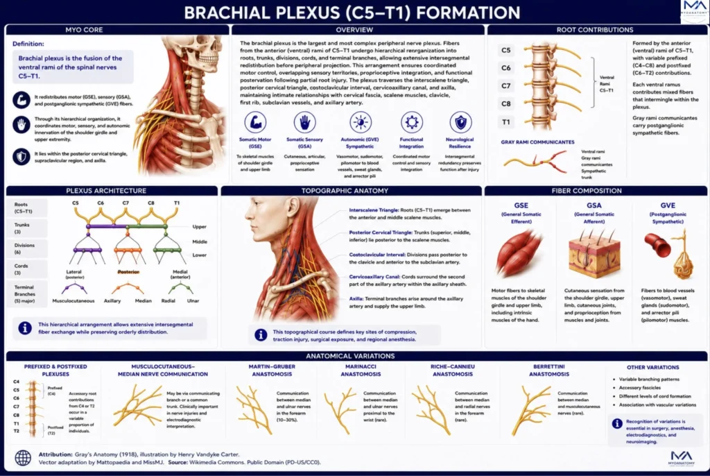
The brachial plexus is the fusion of the anterior (ventral) rami of C5–Th1 spinal nerves. Through its hierarchical organization, it redistributes GSE, GSA, and postganglionic sympathetic GVE fibers to provide integrated motor, sensory, and autonomic innervation of the upper extremity.
OVERVIEW
The brachial plexus is the most complex peripheral nerve plexus, providing the exclusive neural continuity between the cervical spinal cord and the upper limb.
During formation, fibers from the anterior rami of C5–Th1 undergo hierarchical reorganization into roots, trunks, divisions, cords, and terminal branches, enabling extensive intersegmental fiber redistribution before peripheral projection.
This organization ensures coordinated motor control, overlapping sensory territories, proprioceptive integration, and functional preservation following partial root injury.
Extending from the interscalene triangle to the axilla, the plexus maintains close relationships with the cervical fascia, scalene muscles, clavicle, first rib, and major neurovascular structures, forming the anatomical basis for upper-limb biomechanics, peripheral nerve surgery, regional anesthesia, and clinical neuroimaging.
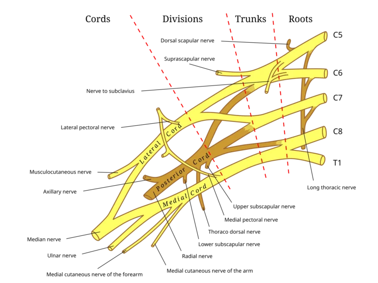
Source: Gray’s Anatomy (1918), illustration by Henry Vandyke Carter. Vector adaptation by Mattopaedia and MissMJ via Wikimedia Commons. Public Domain.
Exam Question
How does the hierarchical organization of the brachial plexus optimize upper-limb function and facilitate clinical localization of neurological lesions?
ANATOMY
Roots Contributions
The brachial plexus is classically formed by the anterior (ventral) rami of C5-Th1, with recognized prefixed (C4-C8) and postfixed (C6-Th2) variants.
Each ventral ramus contributes mixed neural fibers that undergo extensive intersegmental redistribution before peripheral branching, allowing every terminal nerve to contain axons originating from multiple spinal cord segments.
This segmental organization provides functional integration across the upper limb while explaining the predictable patterns of motor and sensory deficits associated with isolated root lesions.
Exam Question
How do normal and variant root contributions determine the segmental organization of the brachial plexus and influence the clinical presentation of root lesions?
Plexus Architecture
The brachial plexus demonstrates a highly ordered hierarchical organization in which 5 roots converge to form 3 trunks, each dividing into anterior and posterior divisions that reorganize into 3 cords, from which the collateral and terminal branches arise.
This sequential arrangement permits extensive intersegmental fiber exchange while maintaining precise anatomical organization for peripheral distribution.
The plexiform architecture provides functional redundancy, coordinated activation of multiple muscle groups, overlapping sensory territories, and accurate anatomical localization of lesions according to the level of plexus involvement.
Exam Question
How does the hierarchical architecture of the brachial plexus facilitate intersegmental fiber redistribution, coordinated upper-limb function, and anatomical localization of neurological lesions?
Topographical Anatomy
The brachial plexus extends from the lower cervical region to the axilla through a continuous series of anatomically confined spaces.
The roots emerge between the anterior and middle scalene muscles within the interscalene triangle, the trunks traverse the posterior cervical triangle, the divisions pass posterior to the clavicle through the costoclavicular space, and the cords enter the cervicoaxillary canal within the axillary sheath surrounding the second part of the axillary artery.
This predictable topographical course defines the principal sites of compression, traction injury, surgical exposure, and regional anesthetic blockade.
Exam Question
How does the topographical course of the brachial plexus influence its vulnerability to compression, traction, and iatrogenic injury throughout the cervicoaxillary region?
Anatomical Relations
Throughout its course, the brachial plexus maintains intimate anatomical relationships with the anterior and middle scalene muscles, prevertebral fascia, first rib, clavicle, subclavian artery, subclavian vein, axillary artery, axillary vein, and the surrounding cervical and axillary musculature.
Distally, the cords are named according to their relationship to the second part of the axillary artery while remaining enclosed within the axillary sheath.
These relationships constitute essential surgical landmarks and underlie the anatomical basis of regional anesthesia, vascular procedures, thoracic outlet pathology, and traumatic neurovascular injuries.
Exam Question
Which anatomical relationships of the brachial plexus are most significant for surgical exposure, regional anesthesia, vascular intervention, and the interpretation of traumatic injuries?
Fiber Composition
The brachial plexus contains general somatic efferent (GSE) fibers supplying the skeletal musculature of the shoulder girdle and upper limb, general somatic afferent (GSA) fibers transmitting cutaneous, proprioceptive, and articular sensation, and postganglionic sympathetic general visceral efferent (GVE) fibers accompanying peripheral nerves to regulate vasomotor tone, sudomotor secretion, and pilomotor activity.
Parasympathetic fibers are not intrinsic components of the brachial plexus, although autonomic pathways traverse the cervicoaxillary region independently of the plexus.
Exam Question
How does the fiber composition of the brachial plexus integrate somatic motor, somatic sensory, and sympathetic autonomic functions throughout the upper extremity?
Anatomical Variations
The brachial plexus exhibits considerable anatomical variation involving root contributions, trunk formation, cord configuration, branching patterns, and neural communications.
Frequent variants include prefixed and postfixed plexuses, musculocutaneous–median nerve communications, and clinically significant interconnections such as the Martin- Gruber, Marinacci, Riche – Cannieu, and Berrettini anastomoses.
Recognition of these variations is essential for peripheral nerve surgery, microsurgical reconstruction, regional anesthesia, electrodiagnostic interpretation, and high-resolution MR neurography, where atypical anatomy may alter both clinical presentation and procedural planning.
Exam Question
Which anatomical variations of the brachial plexus have the greatest implications for peripheral nerve surgery, regional anesthesia, electrodiagnostic interpretation, and advanced neuroimaging, and why?
SUMMARY TABLE