Sacral Plexus
MYO CORE

Formation
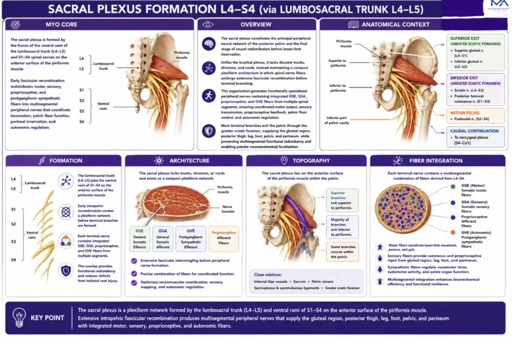
The sacral plexus is a highly organized multisegmental neural network formed by the lumbosacral trunk (L4–L5) and the fusion of the ventral rami of S1-S4 spinal nerves. Through selective division and fiber redistribution, it forms collateral and terminal nerves carrying GSE, GSA, GVE and GVA fibers while preserving segmental organization and the anatomical distinction between anterior and posterior divisions.
OVERVIEW
The sacral plexus constitutes the principal peripheral neural network of the posterior pelvis and the final stage of neural fiber redistribution before lower-limb innervation.
Formed by the lumbosacral trunk (L4–L5) and ventral rami of S1–S4, it lies on the anterior surface of the piriformis muscle.
Unlike the brachial plexus, it does not form trunks, divisions, or cords, but instead maintains a compact plexiform architecture in which extensive intrapelvic fascicular recombination redistributes fibers before terminal nerve formation.
This organization generates multisegmental peripheral nerves containing integrated GSE, GSA, proprioceptive, and postganglionic sympathetic (GVE) fibers, ensuring coordinated motor, sensory, proprioceptive, and autonomic function.
Most terminal branches exit the pelvis through the greater sciatic foramen to supply the gluteal region, posterior thigh, leg, and foot, while pelvic branches remain within the pelvis to innervate the pelvic floor, perineum, and pelvic viscera.
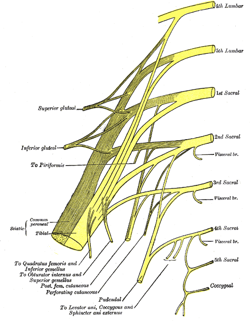
Plan of sacral and pudendal plexuses (Gray828)” – Henry Vandyke Carter, Gray’s Anatomy: Anatomy of the Human Body. Source: Wikimedia Commons. Public Domain.
Exam Question
Explain the formation, anatomical organization, topographical relationships, fiber composition, and functional significance of the sacral plexus, emphasizing how its unique plexiform architecture differs from the brachial plexus and enables coordinated somatic and autonomic innervation of the pelvis and lower limb.
ANATOMY
Roots Contributions
The sacral plexus is formed by the lumbosacral trunk (L4 – L5) together with the ventral rami of S1- S4.
The lumbosacral trunk descends medial to the psoas major, crosses the pelvic brim anterior to the sacroiliac joint and sacral ala, and joins the first sacral ventral ramus on the posterolateral pelvic wall.
Each ventral ramus contributes segmentally organized fascicles that remain anatomically identifiable within the plexus before undergoing selective redistribution into collateral and terminal nerves.
Consequently, every major peripheral branch contains precisely integrated fibers derived from multiple spinal segments while preserving their original segmental topography.
Exam Question
How does the anatomical course of the lumbosacral trunk establish continuity between the lumbar and sacral plexuses, and why is preservation of segmental fascicular topography essential during formation of the sacral plexus?
Topographical Anatomy
The sacral plexus occupies the extraperitoneal compartment of the posterolateral pelvis, resting directly upon the pelvic surface of the piriformis muscle beneath the parietal pelvic fascia.
It extends from the superior margin of the greater sciatic foramen to the pelvic floor and lies in close relationship to the sacral ala, sacroiliac joint, internal iliac artery and its branches, pelvic ureter, rectum, and pelvic autonomic plexuses.
Most collateral and terminal branches converge toward the greater sciatic foramen, where they exit either superior or inferior to the piriformis according to their definitive anatomical distribution.
Exam Question
Discuss how the three-dimensional topographical relationships of the sacral plexus determine the course of its branches and influence surgical approaches to the pelvis and gluteal region.
Plexus Architecture
Within the plexus, each ventral ramus divides into anterior and posterior divisions, which remain anatomically segregated throughout plexus formation.
Rather than forming a random neural meshwork, these divisions undergo highly ordered fascicular recombination, producing peripheral nerves with constant internal organization and reproducible segmental composition.
The anterior divisions predominantly contribute to nerves supplying flexor-derived muscular compartments and pelvic viscera, whereas the posterior divisions contribute principally to extensor-derived muscular territories and the posterior lower limb. This hierarchical fascicular arrangement constitutes the defining architectural feature of the sacral plexus.
Exam Question
Explain how preservation of anterior and posterior divisions determines the internal architecture of the sacral plexus and the segmental composition of its collateral and terminal branches.
Structural Organization
The sacral plexus is best understood as a three-dimensional multisegmental fascicular redistribution system rather than a simple convergence of ventral rami.
Individual fascicles undergo sequential convergence, separation, and recombination while maintaining precise internal topographical orientation throughout the plexus.
This organization preserves predictable relationships between spinal segments and peripheral nerves, facilitates integration between the lumbar and sacral plexuses, and establishes the structural basis for the formation of the sciatic nerve and the remaining collateral and terminal branches.
Exam Question
Why is the sacral plexus regarded as a three-dimensional fascicular redistribution system rather than a simple neural fusion, and how does this organization determine the architecture of its peripheral nerves?
SUMMARY TABLE