Lumbar Plexus
MYO CORE
Clinical Significance
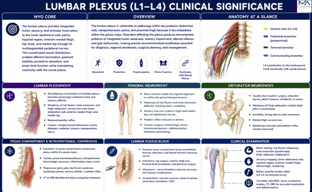
The lumbar plexus occupies a deep retroperitoneal position within the posterior part of the psoas major, making it particularly vulnerable to compression, hemorrhage, infection, neoplastic invasion, surgical trauma, and metabolic neuropathies. Because each peripheral nerve contains overlapping fibers from multiple spinal segments (L1–L4), lesions produce characteristic combinations of motor weakness, sensory impairment, and reflex abnormalities.
OVERVIEW
Lumbar plexus disorders are interpreted by correlating anatomical nerve pathways with patterns of muscle weakness, sensory deficits, proprioceptive impairment, tendon reflex changes, gait abnormalities, and imaging findings.
Lesions may involve the ventral rami, intrapsoas plexus, collateral branches, terminal nerves, or communicating pathways, producing predictable deficits affecting hip flexion, thigh adduction, knee extension, cutaneous sensation, joint stability, and coordinated locomotion.
Comprehensive evaluation integrates neurological examination, muscle testing, dermatomal and peripheral sensory mapping, deep tendon reflexes, electromyography, nerve conduction studies, ultrasound, CT, MRI, and image-guided diagnostic nerve blocks.
Detailed knowledge of lumbar plexus anatomy is therefore fundamental for orthopedic surgery, pelvic and retroperitoneal procedures, regional anesthesia, trauma care, neurology, rehabilitation, and pain medicine.
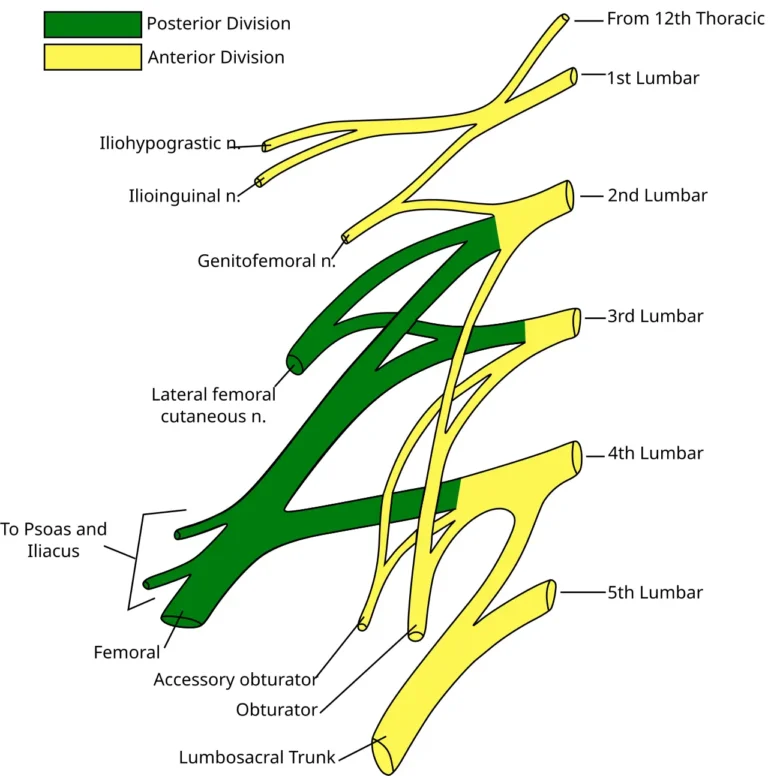
Lumbar Plexus. Adapted from Gray822_es.svg by Gray; derivative work by Ninovolador and Mcstrother. Licensed under CC BY 3.0 via Wikimedia Commons.
Exam Question
How does the lumbar plexus integrate multisegmental spinal output into coordinated peripheral innervation that supports posture, locomotion, proprioception, and lower-limb biomechanics?
ANATOMY
Lumbar Plexopathy
Lumbar plexopathy is characterized by simultaneous involvement of multiple branches of the lumbar plexus, producing neurological deficits that extend beyond the distribution of a single peripheral nerve or spinal root.
Patients commonly develop weakness of hip flexion, knee extension, and thigh adduction with sensory loss involving the lower abdominal wall, inguinal region, anterior-medial thigh, medial leg, and variable depression or absence of the patellar reflex.
Frequent etiologies include retroperitoneal hematoma, diabetic lumbosacral radiculoplexus neuropathy, pelvic malignancy, radiation-induced fibrosis, traumatic injury, postoperative complications, and inflammatory plexitis. Recognition of this mixed motor-sensory pattern is essential for differentiating plexopathy from isolated radiculopathy or mononeuropathy.
Exam Question
How do the combined patterns of motor weakness, sensory impairment, reflex abnormalities, and electrodiagnostic findings distinguish lumbar plexopathy from L2–L4 radiculopathy, isolated femoral neuropathy, obturator neuropathy, and upper motor neuron disorders?
Femoral Neuropathy
Femoral neuropathy results in dysfunction of the quadriceps femoris and variable involvement of the iliopsoas, producing weakness of hip flexion, loss of knee extension, difficulty climbing stairs, impaired sit-to-stand transition, and instability during the stance phase of gait.
Sensory loss affects the anterior thigh and medial leg through the saphenous nerve, while the patellar tendon reflex is reduced or absent.
Common causes include pelvic surgery, femoral artery catheterization, retroperitoneal hemorrhage, psoas pathology, inguinal compression, and pelvic fractures. Persistent denervation leads to quadriceps atrophy and significant impairment of lower-limb biomechanics.
Exam Question
How does the anatomical course of the femoral nerve explain the characteristic combination of quadriceps weakness, diminished patellar reflex, anterior thigh and medial leg sensory loss, and gait instability following retroperitoneal, pelvic, or inguinal pathology?
Obturator Neuropathy
Obturator nerve injury primarily affects the adductor compartment of the thigh, causing weakness of thigh adduction, impaired pelvic stabilization during gait, reduced balance during directional changes, and difficulty crossing the legs.
Patients frequently report medial thigh pain radiating toward the knee together with sensory loss over the distal medial thigh, whereas quadriceps strength and the patellar reflex usually remain preserved.
Injury most commonly occurs following pelvic fractures, obturator hernia, pelvic lymph-node dissection, gynecological surgery, childbirth, pelvic tumors, or chronic athletic overuse.
Exam Question
Why does isolated obturator neuropathy produce selective impairment of thigh adduction and dynamic pelvic stabilization while preserving knee extension and the patellar tendon reflex, and how can it be differentiated clinically from femoral neuropathy and upper lumbar radiculopathy?
Psoas Compartment
The lumbar plexus forms within the posterior third of the psoas major, making it highly susceptible to mass effect from retroperitoneal pathology.
Psoas hematoma, abscess, retroperitoneal hemorrhage, abdominal aortic aneurysm, enlarged lymph nodes, metastatic disease, and primary retroperitoneal tumors may simultaneously compress multiple lumbar nerves, producing groin pain, hip flexion weakness, quadriceps paralysis, sensory deficits, and loss of the patellar reflex. CT and MRI accurately define lesion extent, while ultrasound assists in selected interventional procedures and drainage.
Exam Question
How does the intimate anatomical relationship between the lumbar plexus and the psoas major explain the simultaneous involvement of multiple peripheral nerves in retroperitoneal disease, and which neurological findings most reliably localize compression to the psoas compartment?
Lumbar Plexus Block
The lumbar plexus (posterior psoas compartment) block is an advanced regional anesthetic technique in which local anesthetic is deposited adjacent to the plexus within the psoas major under ultrasound guidance and/or peripheral nerve stimulation.
It provides reliable anesthesia of the femoral, obturator, and lateral femoral cutaneous nerves for hip arthroplasty, femoral fracture fixation, anterior thigh surgery, and selected knee procedures.
Precise anatomical localization minimizes complications including epidural spread, retroperitoneal hematoma, renal puncture, vascular injury, nerve trauma, and local anesthetic systemic toxicity.
Exam Question
How do the three-dimensional anatomy of the lumbar plexus within the psoas compartment and the fascial relationships of surrounding structures determine the effectiveness, sensory distribution, motor blockade, and potential complications of a lumbar plexus block?
Clinical Examination
Clinical localization of lumbar plexus lesions integrates motor testing, sensory mapping, tendon reflexes, gait assessment, and electrodiagnostic evaluation.
Hip flexion, knee extension, and thigh adduction assess the principal motor branches, while sensory examination includes the anterior and medial thigh and medial leg. The patellar reflex evaluates femoral nerve integrity and L2 – L4 segments.
Electromyography, nerve conduction studies, and MRI or CT confirm lesion localization and differentiate lumbar plexopathy from radiculopathy and isolated peripheral neuropathies.
Exam Question
How do motor testing, sensory mapping, tendon reflexes, and electrodiagnostic studies accurately localize lumbar plexus lesions and distinguish them from radiculopathy and isolated peripheral neuropathies?
SUMMARY TABLE