Cervical Plexus
MYO CORE
Formation
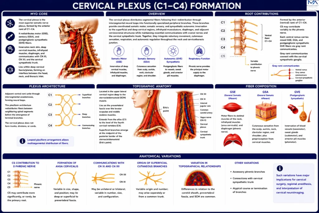
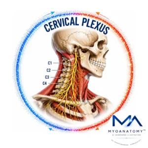
The cervical plexus formed by fusion of the anterior (ventral) rami of C1–C4 spinal nerves. Through segmental neural interconnections, it redistributes general somatic efferent (GSE), general somatic afferent (GSA), and postganglionic sympathetic general visceral efferent (GVE) fibers into cutaneous, muscular, and communicating branches.
OVERVIEW
The cervical plexus represents the principal somatic neural network of the cervical region.
It is established through intercommunication of the anterior rami of C1 – C4, producing a plexiform arrangement that redistributes motor, sensory, and autonomic fibers before peripheral distribution.
This organization provides multisegmental innervation to the superficial cervical integument, deep cervical musculature, infrahyoid muscles, and diaphragm.
Meanwhile to that, cervical plexus maintaining functional communications with the hypoglossal nerve (CN XII), accessory nerve (CN XI), cervical sympathetic trunk, and adjacent cervical neurovascular structures.
Its position within the deep cervical fascial planes establishes an important anatomical interface between the head, neck, and thoracic inlet.
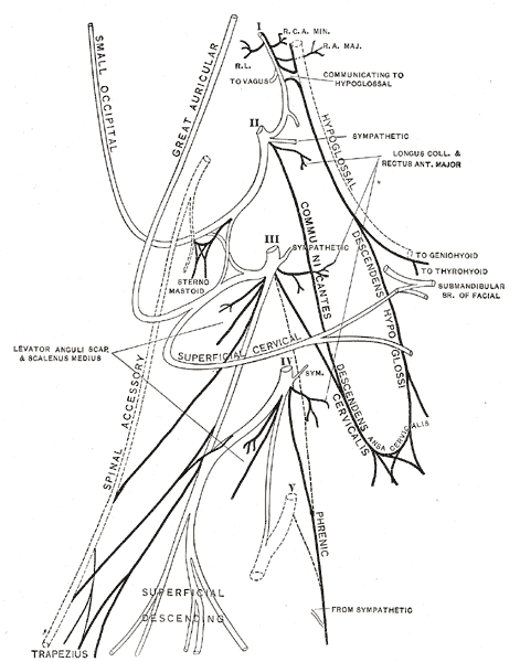
Henry Gray, Anatomy of the Human Body (20th ed., 1918), Plate 804. Illustration by Henry Vandyke Carter via Wikimedia Commons. Public Domain.
Exam Question
How does the multisegmental organization of the cervical plexus optimize the integration of motor, sensory, and autonomic innervation within the cervical region?
ANATOMY
Roots Contributions
The cervical plexus is formed predominantly by the anterior (ventral) rami of the C1–C4 spinal nerves, with occasional contribution from C5 to the phrenic nerve.
Following their emergence through the corresponding intervertebral foramina, each ventral ramus carries mixed general somatic efferent (GSE), general somatic afferent (GSA), and postganglionic sympathetic general visceral efferent (GVE) fibers received through the gray rami communicantes of the cervical sympathetic trunk.
These fibers subsequently intermingle within the plexus before being redistributed into its peripheral branches, establishing overlapping segmental innervation throughout the cervical region.
Exam Question
How do the segmental contributions of C1–C4, together with the variable C5 contribution to the phrenic nerve, establish the functional organization, segmental overlap, and clinical variability of the cervical plexus?
Plexus Architecture
The cervical plexus is organized as a series of intersegmental neural loops formed by adjacent anastomoses between the anterior rami of C1–C4.
This plexiform architecture permits redistribution of neural fibers between neighboring spinal segments before the emergence of terminal branches, ensuring coordinated multisegmental motor, sensory, and autonomic innervation.
Unlike the brachial plexus, the cervical plexus retains a relatively simple looped configuration and does not differentiate into trunks, divisions, or cords, reflecting its predominantly regional pattern of innervation.
Exam Question
How does the looped intersegmental architecture of the cervical plexus facilitate neural fiber redistribution, and why does this organization differ fundamentally from the trunk–division–cord arrangement of the brachial plexus?
Topographical Anatomy
The cervical plexus occupies the upper lateral cervical region within the posterior part of the carotid triangle, lying deep to the sternocleidomastoid muscle and enclosed by the prevertebral layer of the deep cervical fascia.
It rests upon the levator scapulae, middle scalene, and adjacent prevertebral muscles, extending from the level of the atlas (C1) to approximately the fourth cervical vertebra (C4).
Its superficial cutaneous branches converge at the midpoint of the posterior border of the sternocleidomastoid, the nerve point of the neck (Erb’s point), where they emerge from the deep fascia to enter the superficial tissues.
Exam Question
How does the topographic position of the cervical plexus within the lateral cervical region and deep cervical fascial compartments determine its anatomical accessibility, surface landmarks, and regional distribution?
Anatomical Relations
The cervical plexus maintains intimate anatomical relationships with the surrounding muscular, fascial, neurovascular, and neural structures of the neck.
Superficially, it is covered by the sternocleidomastoid muscle and the investing layer of the deep cervical fascia.
Medially, it lies adjacent to the carotid sheath, containing the common and internal carotid arteries, internal jugular vein, and vagus nerve (CN X).
Posteriorly, it is supported by the levator scapulae, middle scalene, splenius capitis, and longus capitis muscles.
Superiorly, it communicates with the hypoglossal nerve (CN XII) and accessory nerve (CN XI), while postganglionic sympathetic fibers reach the plexus through the cervical sympathetic trunk. These spatial relationships define important surgical planes and anatomical landmarks for cervical dissection and regional anesthesia.
Exam Question
Which muscular, fascial, neurovascular, and neural relationships are most critical to the cervical plexus, and how do these spatial relationships influence surgical approaches, regional anesthesia, and the risk of iatrogenic injury?
Fiber Compositions
The cervical plexus contains three principal functional fiber modalities.
General somatic efferent (GSE) fibers provide motor innervation to skeletal muscles of the cervical region, including the infrahyoid musculature through the ansa cervicalis and the diaphragm through the phrenic nerve.
General somatic afferent (GSA) fibers transmit cutaneous sensation from the anterolateral neck, auricle, occipital region, clavicular region, and superior shoulder, together with proprioceptive information from the cervical musculature.
Postganglionic sympathetic general visceral efferent (GVE) fibers accompany the peripheral branches to innervate vascular smooth muscle, sweat glands, and arrector pili muscles, thereby regulating vasomotor, sudomotor, and pilomotor functions throughout their distribution.
Exam Question
How do GSE, GSA, and postganglionic sympathetic GVE fibers integrate within the cervical plexus to produce coordinated motor, sensory, and autonomic function throughout its peripheral distribution?
Anatomical Variations
The cervical plexus demonstrates considerable variation in its segmental contributions, intersegmental communications, branching patterns, and topographical relationships.
The most frequent variations involve the contribution of C5 to the phrenic nerve, the configuration of the ansa cervicalis, communications with the hypoglossal nerve (CN XII) and accessory nerve (CN XI), and the origin, course, and distribution of the superficial cutaneous branches.
Less commonly, variations occur in the relationship of the plexus to the sternocleidomastoid muscle, carotid sheath, and prevertebral fascia. Recognition of these anatomical variants is essential for accurate surgical dissection, regional anesthesia, and radiological interpretation.
Exam Question
Which anatomical variations of the cervical plexus most significantly influence cervical surgery, regional anesthesia, and radiological interpretation, and what are their underlying anatomical mechanisms?
SUMMARY TABLE