Cervical Plexus
MYO CORE
Clinical Significance
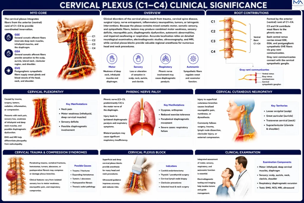
The cervical plexus is a clinically significant neural network whose integrity is essential for normal cervical sensation, neck muscle function, swallowing, and diaphragmatic respiration. Because of its superficial location, extensive communications with cranial nerves, and close relationship to major cervical neurovascular structures, it is particularly vulnerable to traumatic injury, surgical intervention, compressive pathology, and regional anesthesia.
OVERVIEW
Disorders of the cervical plexus encompass a broad spectrum of traumatic, compressive, inflammatory, neoplastic, vascular, and iatrogenic conditions affecting its peripheral branches.
Because the plexus contributes to cervical motor control, cutaneous sensation, diaphragmatic function, and regional neural integration, pathology may produce diverse clinical manifestations ranging from localized sensory disturbances and neck pain to diaphragmatic paralysis, dysphagia, respiratory compromise, and complex neurological deficits.
Comprehensive evaluation requires correlation of anatomical findings with neurological examination, electrodiagnostic testing, ultrasonography, and cross-sectional imaging to accurately localize lesions, differentiate plexopathy from adjacent neurological disorders, and guide appropriate medical, surgical, or anesthetic management.
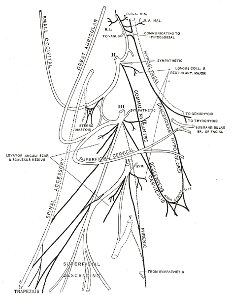
Henry Gray, Anatomy of the Human Body (20th ed., 1918), Plate 804. Illustration by Henry Vandyke Carter via Wikimedia Commons. Public Domain.
Exam Question
How do the anatomical organization, functional distribution, and neurovascular relationships of the cervical plexus determine the clinical presentation, diagnostic localization, and management of cervical plexus disorders?
ANATOMY
Cervical Plexopathy
Cervical plexopathy is an uncommon disorder resulting from traumatic injury, cervical surgery, neoplastic infiltration, radiation-induced fibrosis, inflammatory neuropathies, or compressive lesions involving multiple branches of the plexus.
Simultaneous involvement of motor and sensory fibers produces varying combinations of cervical pain, sensory loss over the lateral neck and shoulder, weakness of the infrahyoid and deep cervical musculature, and diaphragmatic dysfunction secondary to phrenic nerve involvement.
Electrodiagnostic studies combined with cervical MRI are fundamental for differentiating cervical plexopathy from cervical radiculopathy, brachial plexopathy, and isolated peripheral neuropathies.
Exam Question
How do the anatomical distribution and functional organization of the cervical plexus explain the neurological deficits of cervical plexopathy, and differentiate it from cervical radiculopathy and brachial plexopathy?
Phrenic Nerve Palsy
The phrenic nerve (C3–C5, predominantly C4) provides the exclusive motor innervation of the diaphragm and constitutes the principal respiratory branch of the cervical plexus.
Injury may occur during cervical, cardiothoracic, or mediastinal surgery, penetrating trauma, regional anesthesia, tumor infiltration, or inflammatory neuropathy.
Clinical manifestations include ipsilateral diaphragmatic paralysis, exertional dyspnea, orthopnea, reduced ventilatory capacity, and paradoxical diaphragmatic elevation during inspiration. Bilateral involvement may result in severe respiratory insufficiency requiring ventilatory support.
Exam Question
How do the anatomical course and functional role of the phrenic nerve explain the respiratory manifestations of phrenic nerve palsy, and why is lesion localization clinically important?
Cervical Cutaneous Neuropathy
Injury to the lesser occipital, great auricular, transverse cervical, or supraclavicular nerves produces localized neuropathic pain, hypoesthesia, dysesthesia, or paresthesia confined to their respective cutaneous territories.
These neuropathies most commonly occur following cervical lymph node dissection, carotid endarterectomy, parotid surgery, thyroid surgery, clavicular trauma, or prolonged external compression. Precise recognition of the sensory deficit assists in localization of the affected branch.
Exam Question
How does the cutaneous distribution of the superficial cervical plexus branches enable precise localization of cervical cutaneous neuropathies, and which surgical procedures place these nerves at greatest risk?
Cervical Trauma
Penetrating cervical trauma, vertebral fractures, expanding hematomas, deep neck infections, tumors, postoperative fibrosis, and other space-occupying lesions may compress or disrupt individual branches or the cervical plexus as a whole.
Clinical manifestations depend on the anatomical level and extent of involvement, ranging from isolated sensory deficits to complex motor weakness, chronic neuropathic pain, dysphagia, diaphragmatic dysfunction, and respiratory compromise associated with phrenic nerve injury.
Exam Question
How do the anatomical relationships of the cervical plexus determine the mechanisms, clinical manifestations, and severity of traumatic and compressive injuries?
Cervical Plexus Block
Superficial and deep cervical plexus blocks are established regional anesthetic techniques employed for carotid endarterectomy, thyroidectomy, parathyroidectomy, cervical lymph node biopsy, clavicular procedures, and selected head and neck operations.
Ultrasound-guided administration improves anatomical accuracy, increases block success, and minimizes complications including inadvertent intravascular injection, neuraxial spread, recurrent laryngeal nerve blockade, and phrenic nerve paralysis.
Exam Question
How does detailed knowledge of cervical plexus anatomy optimize the efficacy and safety of superficial and deep cervical plexus blocks while minimizing procedure-related complications?
Clinical Examination
Evaluation of cervical plexus function requires integrated assessment of motor, sensory, respiratory, and autonomic function.
Motor examination includes the infrahyoid musculature, deep cervical flexors, and diaphragmatic performance, whereas sensory testing evaluates the cutaneous territories of the lesser occipital, great auricular, transverse cervical, and supraclavicular nerves.
Diaphragmatic function is assessed by ultrasonography or fluoroscopy, while electromyography, nerve conduction studies, and cervical MRI facilitate lesion localization and differentiation from cervical radiculopathy, brachial plexopathy, or isolated peripheral nerve disorders.
Exam Question
How does systematic motor, sensory, respiratory, and electrodiagnostic assessment accurately localize cervical plexus lesions and distinguish them from other peripheral nerve disorders?
SUMMARY TABLE