Cervical Plexus
MYO CORE
Branches
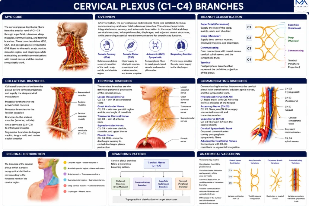
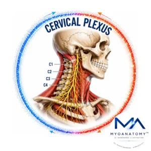
The cervical plexus formed by fusion of (ventral) rami of C1–C4 of spinal nerves through an organized network of cutaneous, muscular, communicating, and terminal branches. These branches integrate general somatic efferent (GSE), general somatic afferent (GSA), and postganglionic sympathetic general visceral efferent (GVE) fibers to establish coordinated motor, sensory, and autonomic innervation throughout the cervical region, diaphragm, and adjacent cervicocranial structures.
OVERVIEW
Following formation of its intersegmental neural network, the cervical plexus undergoes orderly peripheral branching that redistributes segmental nerve fibers into anatomically and functionally specialized branches.
These branches are organized according to their anatomical origin, topographical course, functional composition, and peripheral distribution as cutaneous, muscular, communicating, and terminal branches.
Collectively, they constitute the principal neural network of the cervical region, providing integrated innervation to the superficial cervical integument, deep cervical musculature, infrahyoid muscles, diaphragm, and adjacent cervicocranial structures while maintaining essential communications with the hypoglossal nerve (CN XII), accessory nerve (CN XI), vagus nerve (CN X), and cervical sympathetic trunk.
This hierarchical branching architecture ensures coordinated segmental transmission of somatic motor, somatic sensory, and autonomic fibers throughout the neck and cervicothoracic junction.
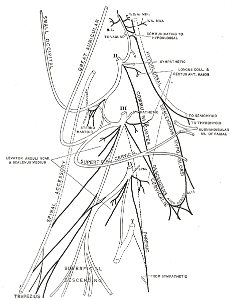
Henry Gray, Anatomy of the Human Body (20th ed., 1918), Plate 804. Illustration by Henry Vandyke Carter via Wikimedia Commons. Public Domain.
Exam Question
How does the hierarchical branching architecture of the cervical plexus facilitate coordinated segmental redistribution of GSE, GSA, and postganglionic sympathetic GVE fibers while preserving functional integration throughout the cervical region?
ANATOMY
Branch Classification
The branches of the cervical plexus are organized according to their segmental derivation, topographical emergence, fascial relationships, functional fiber modalities, and peripheral projection.
Following intersegmental redistribution within the plexiform network, fibers originating from the anterior (ventral) rami of C1–C4 segregate into cutaneous, muscular, communicating, and terminal branches, each representing a specialized anatomical pathway for the transmission of general somatic efferent (GSE), general somatic afferent (GSA), and accompanying postganglionic sympathetic general visceral efferent (GVE) fibers.
This hierarchical branching pattern preserves segmental overlap, enhances functional integration, and establishes the structural framework through which cervical spinal nerves coordinate motor, sensory, and autonomic innervation throughout the neck.
Exam Question
How does hierarchical branch classification reflect the structural organization, segmental redistribution, and functional specialization of fibers within the cervical plexus following formation of its intersegmental neural network?
Collateral Branches
Collateral branches arise directly from the cervical plexus before terminal peripheral projection and constitute the primary motor distribution to the deep cervical musculature.
These branches supply the longus capitis, longus colli, rectus capitis anterior, rectus capitis lateralis, levator scapulae, and portions of the scalene musculature, while fibers from C1–C3 contribute to formation of the ansa cervicalis, which provides motor innervation to the majority of the infrahyoid muscles.
Through their intimate relationship with the prevertebral fascia and cervical muscular compartments, collateral branches integrate cervical posture, craniocervical stability, swallowing, respiratory mechanics, and proprioceptive feedback, while preserving overlapping multisegmental motor control.
Exam Question
How do the collateral branches integrate segmental motor innervation of the deep cervical musculature and infrahyoid compartment while preserving coordinated cervical stability, swallowing, respiration, and proprioceptive function?
Terminal Branches
The terminal branches represent the definitive peripheral projection of cervical plexus fibers and include the lesser occipital, great auricular, transverse cervical, supraclavicular, and phrenic nerves. Their emergence reflects the final redistribution of fibers following plexus formation, allowing precise delivery of sensory, motor, and accompanying sympathetic fibers to their target tissues.
The superficial branches provide cutaneous innervation to the scalp, auricle, parotid region, anterior and lateral neck, clavicular region, and shoulder, whereas the phrenic nerve constitutes the principal motor pathway to the diaphragm while also conveying sensory fibers from the central diaphragm, fibrous pericardium, and mediastinal and diaphragmatic pleura.
Together, these branches establish the functional interface between cervical spinal segments and peripheral cervical, thoracic, and respiratory structures.
Exam Question
How do the anatomical origin, topographical course, and functional specialization of each terminal branch reflect the segmental architecture and peripheral projection of fibers derived from the cervical plexus?
Communicating Branches
Communicating branches establish complex neural interconnections between the cervical plexus and the hypoglossal nerve (CN XII), accessory nerve (CN XI), vagus nerve (CN X), the cervical sympathetic trunk, and adjacent cervical spinal nerves.
These communications facilitate the exchange of somatic motor, proprioceptive, and autonomic fibers, ensuring coordinated activity between spinal and cranial neural systems. The superior root of the ansa cervicalis, formed by C1 fibers traveling with the hypoglossal nerve, exemplifies this functional integration.
Collectively, these communicating pathways maintain synchronized cervical motor control, autonomic regulation, and neurovascular coordination throughout the head and neck.
Exam Question
How do the communicating branches function as neuroanatomical interfaces integrating spinal, cranial, and sympathetic pathways to coordinate motor, sensory, proprioceptive, and autonomic function within the cervicocranial region?
Regional Distribution
The branches of the cervical plexus demonstrate a highly organized topographical distribution corresponding to the anatomical compartments of the neck.
Superficial branches supply the occipital region, auricle, parotid region, anterolateral cervical integument, supraclavicular region, and superior shoulder, whereas deep branches innervate the prevertebral muscles, infrahyoid musculature, and diaphragm.
This compartmentalized organization enables coordinated integration of cutaneous sensation, skeletal motor function, proprioception, respiration, and sympathetic regulation while maintaining continuity across the superficial and deep fascial planes of the cervical region.
Exam Question
How does the compartmental topographical distribution of cervical plexus branches determine functional integration of the superficial neck, deep cervical musculature, shoulder girdle, diaphragm, and adjacent cervicothoracic structures?
Branching Pattern
The branching pattern of the cervical plexus reflects the sequential organization of its segmental fiber redistribution following formation of the plexiform network.
Superficial cutaneous branches emerge collectively at the nerve point of the neck (Erb’s point) before diverging toward their respective cutaneous territories, whereas deep muscular branches remain within the prevertebral compartment to supply the cervical musculature and contribute to the ansa cervicalis and phrenic nerve.
The arrangement, caliber, and trajectory of individual branches demonstrate predictable anatomical organization while preserving sufficient variability to maintain overlapping segmental innervation and functional redundancy throughout the cervical region.
Exam Question
How does the branching pattern of the cervical plexus reflect the sequential redistribution of segmental fibers, and how does this organization optimize functional innervation of the superficial and deep cervical compartments?
SUMMARY TABLE