Venous Drainage

MYO CORE

Structural Organization

The venous system is organized as a hierarchically integrated, high-compliance vascular network forming the low-pressure outflow component of systemic circulation

OVERVIEW

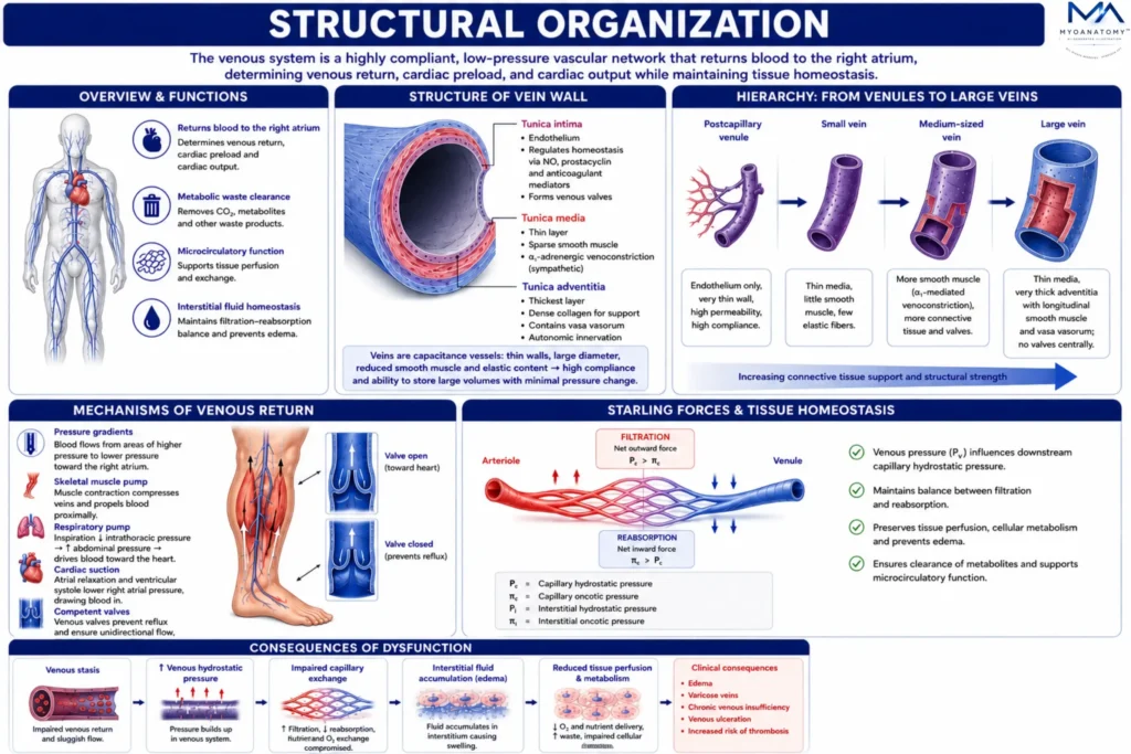
The venous system is a highly compliant, low-pressure vascular network that returns blood to the right atrium, determining venous return, cardiac preload, and cardiac output while maintaining metabolic waste clearance, microcirculatory function, and interstitial fluid homeostasis.

Veins are specialized capacitance vessels with thin walls, reduced smooth muscle and elastic content, and large luminal diameter, allowing high-volume storage with minimal pressure change. The endothelium regulates vascular homeostasis through nitric oxide, prostacyclin, and anticoagulant mediators, while venous valves ensure unidirectional flow and prevent reflux.

The tunica media contains sparse smooth muscle mediating α₁-adrenergic venoconstriction, whereas the tunica adventitia provides structural support, vasa vasorum, and autonomic innervation. Veins are organized from postcapillary venules to large veins, with increasing connective tissue support.

Venous return is maintained by pressure gradients, the skeletal muscle pump, respiratory pump, cardiac suction, and competent valves. Venous pressure regulates Starling forces, preserving filtration-reabsorption balance, tissue perfusion, cellular metabolism, and preventing edema. Dysfunction causes venous stasis, elevated hydrostatic pressure, impaired capillary exchange, and interstitial fluid accumulation.

Exam Question

How do the structural and functional adaptations of the venous system collectively regulate venous return, cardiac preload, Starling forces, and tissue fluid homeostasis, and what are the physiological consequences of their dysfunction?

ANATOMY

Venules

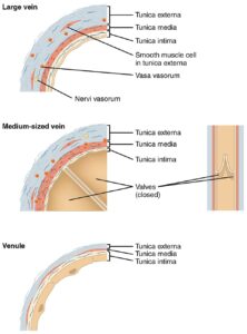
Venules are the initial venous vessels receiving blood from capillary beds and connecting the microcirculation to systemic venous return. Beyond drainage, they regulate capillary pressure, vascular permeability, leukocyte trafficking, and microvascular homeostasis.

Their thin endothelial walls, with minimal or absent tunica media and sparse connective tissue, provide high compliance and permeability, facilitating transendothelial fluid exchange and serving as the principal site of leukocyte adhesion and extravasation during inflammation.

Venules also regulate Starling forces by controlling downstream venous pressure, maintaining the balance between capillary filtration and reabsorption.

In musculoskeletal tissues, they promote metabolite clearance, immune cell recruitment, and tissue repair during mechanical loading, hypoxia, and injury.

Large Vein, Medium Vein, and Venule (Histological Structure)” – OpenStax College via Wikimedia Commons. Licensed under CC BY 3.0

Exam Question

How do the structural characteristics of venules enable regulation of microvascular permeability, leukocyte extravasation, and Starling forces during inflammation and tissue repair?

Mid-sized Veins

Medium-sized veins are the principal conduits of venous return, transporting blood from venules to larger veins while regulating venous tone, capacitance, and flow distribution under low-pressure conditions.

Their trilaminar wall includes a thin tunica media with α₁-adrenergic-responsive smooth muscle that mediates venoconstriction and central blood volume redistribution.

The tunica adventitia, the dominant layer, provides structural support and contains vasa vasorum and autonomic nerves.

Venous valves, formed by endothelial folds reinforced with connective tissue, prevent reflux and, together with the skeletal muscle pump, generate pressure gradients that overcome gravity and maintain unidirectional flow.

Within the musculoskeletal system, medium-sized veins travel within neurovascular bundles, coordinating venous return with arterial perfusion and neural regulation to sustain metabolic exchange and hemodynamic stability during posture and muscle activity.

Large Vein, Medium Vein, and Venule (Histological Structure)” – OpenStax College via Wikimedia Commons. Licensed under CC BY 3.0

Exam Question

How do the structural adaptations of medium-sized veins optimize venous return, regulate capacitance, and maintain unidirectional blood flow during postural change and skeletal muscle contraction?

Large Veins

Large veins are the terminal collecting vessels that return blood to the right atrium, determining venous return, cardiac preload, and cardiac output while serving as the body’s principal capacitance reservoirs.

They possess a thin tunica media and a dominant tunica adventitia containing dense collagen, longitudinal smooth muscle, vasa vasorum, and autonomic nerves, providing structural support, maintaining vessel patency, and regulating venous tone.

The tunica intima forms a continuous endothelium, whereas valves are generally absent in central veins because of their proximity to the heart.

Their high compliance permits storage and rapid mobilization of blood during sympathetic venoconstriction, increasing venous return and cardiac preload.

Blood flow is driven by pressure gradients, respiratory mechanics, and cardiac suction, while the large lumen minimizes vascular resistance. Ultimately, large veins converge into the superior and inferior vena cava, ensuring continuous venous return and central hemodynamic stability.

Large Vein, Medium Vein, and Venule (Histological Structure)” – OpenStax College via Wikimedia Commons. Licensed under CC BY 3.0

Exam Question

How do the structural adaptations of large veins optimize venous capacitance, regulate cardiac preload, and maintain central hemodynamic stability?

SUMMARY TABLE

Scroll to Top