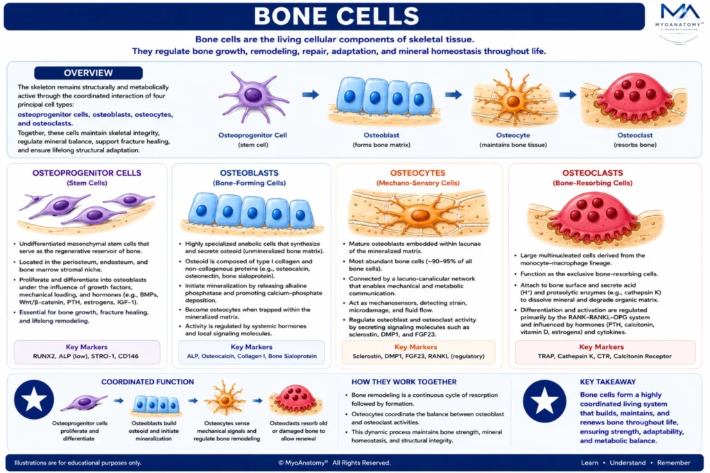
Bone Architecture
MYO CORE
Bone Cells
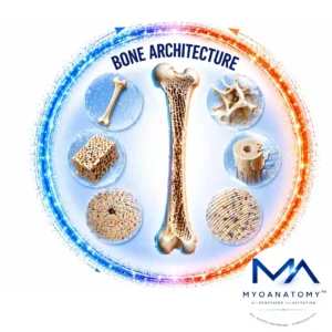
Bone cells constitute a coordinated cellular network that regulates skeletal growth, bone formation, remodeling, repair, and mineral homeostasis. Through the integrated activity of osteoprogenitor cells, osteoblasts, osteocytes, and osteoclasts, bone maintains structural integrity, adapts to mechanical stress, and undergoes continuous lifelong renewal.
OVERVIEW
Osteoprogenitor cells constitute the regenerative stem-cell population of skeletal tissue and serve as the principal cellular reservoir for bone growth, repair, and remodeling. Through tightly regulated differentiation pathways, these precursor cells give rise to osteoblasts, which synthesize osteoid and initiate the mineralization process responsible for the formation of new bone matrix.
Osteocytes, derived from osteoblasts that become embedded within the mineralized extracellular matrix, represent the dominant cellular population of mature bone. Through their extensive lacuno-canalicular communication network, they function as the primary mechanosensory and regulatory system of the skeleton, continuously monitoring mechanical strain, microdamage, fluid dynamics, and metabolic demands while coordinating adaptive skeletal responses.
Osteoclasts are highly specialized multinucleated cells responsible for controlled bone resorption. By removing aged, damaged, or mechanically inefficient bone tissue, they maintain skeletal quality, facilitate mineral mobilization, and create the structural foundation necessary for subsequent bone formation and renewal.
Together, osteoprogenitor cells, osteoblasts, osteocytes, and osteoclasts form a highly integrated cellular network that governs skeletal growth, mineral homeostasis, fracture repair, biomechanical adaptation, and lifelong tissue renewal.
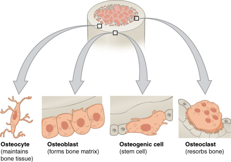
“Bone cells” – OpenStax College, Anatomy & Physiology, Connexions Web site via Wikimedia Commons. Licensed under CC BY 3.0.
ANATOMY
Osteogenic Potential
Osteoprogenitor cells constitute the undifferentiated mesenchymal stem-cell population of skeletal tissue and serve as the principal regenerative reservoir of bone.
Located within the periosteum and endosteum, these precursor cells proliferate and differentiate into osteoblasts under the influence of local growth factors, mechanical loading, and systemic hormones including growth hormone, thyroid hormones, estrogen, testosterone, and parathyroid hormone.
Through this hormonally regulated differentiation pathway, osteoprogenitor cells provide the cellular basis for skeletal growth, fracture healing, remodeling, and lifelong regenerative capacity.
Exam Question
How do osteoprogenitor cells integrate local growth factors, mechanical stimuli, and systemic hormonal regulation to maintain the regenerative capacity of skeletal tissue, while simultaneously supporting bone growth, fracture healing, remodeling, and lifelong skeletal renewal?
Matrix Synthesis
Osteoblasts cells are highly specialized anabolic cells responsible for the production, organization, and mineralization of bone matrix. They synthesize osteoid, a collagen-rich extracellular framework that subsequently mineralizes through calcium-phosphate deposition to form mature skeletal tissue.
Osteoblastic activity is regulated by multiple signaling pathways, including BMP, Wnt/β-catenin, IGF-1, and PTH-mediated mechanisms, allowing bone formation to respond to developmental, hormonal, and mechanical demands.
Beyond matrix production, osteoblasts regulate osteoclast differentiation and coordinate the formation phase of skeletal remodeling.
Exam Question
How do osteoblasts coordinate osteoid synthesis, extracellular matrix organization, mineral deposition, and osteoclast regulation to achieve controlled skeletal formation and maintain structural adaptation in response to developmental, hormonal, and mechanical demands?
Mechanosensory Network
Osteocytes are terminally differentiated osteoblasts embedded within the mineralized matrix and represent the dominant cellular population of bone. Through an extensive lacuno-canalicular communication network, they function as the primary mechanosensory and endocrine-regulatory system of the skeleton.
Osteocytes continuously detect mechanical strain, microdamage, fluid movement, and metabolic alterations, converting these stimuli into molecular signals that regulate both osteoblastic bone formation and osteoclastic resorption.
By secreting signaling molecules such as sclerostin, RANKL, nitric oxide, and prostaglandins, osteocytes integrate hormonal, mechanical, and metabolic information to maintain skeletal homeostasis and structural adaptation.
Exam Question
How does the osteocytic lacuno-canalicular network function as the principal mechanosensory and endocrine-regulatory system of bone, enabling the detection of mechanical strain, microdamage, and metabolic alterations while coordinating skeletal homeostasis and structural adaptation?
Remodeling Dynamics
Osteoclasts are large multinucleated cells derived from the monocyte-macrophage lineage and function as the exclusive resorptive cells of bone.
Their differentiation and activity are primarily regulated through the RANK-RANKL-OPG signaling system and are strongly influenced by parathyroid hormone, calcitonin, vitamin D, estrogen, and inflammatory cytokines. Osteoclasts create a specialized resorption compartment where hydrogen ions and proteolytic enzymes dissolve mineralized matrix and degrade organic bone tissue.
This precisely controlled process removes aged or damaged bone, releases stored minerals into circulation, and permits replacement by newly formed bone, thereby ensuring continuous skeletal renewal, biomechanical optimization, and mineral homeostasis throughout life.
Exam Question
How does RANK-RANKL-OPG-mediated osteoclast regulation control bone resorption, mineral homeostasis, and skeletal renewal, and why is precise coordination between osteoclastic resorption and osteoblastic formation essential for long-term biomechanical integrity?
SUMMARY TABLE