Bone Remodeling

MYO CORE

Remodeling Cycle

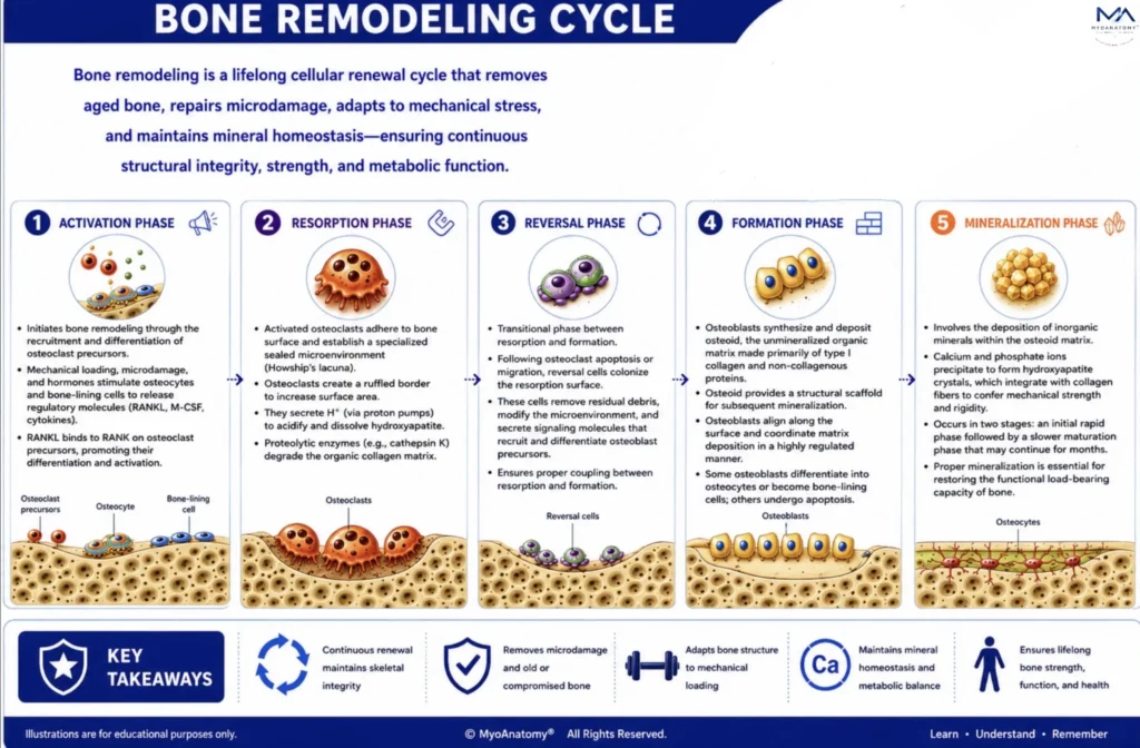
Bone remodeling is a lifelong cellular renewal cycle that removes aged bone, repairs microdamage, adapts skeletal architecture to mechanical stress, and maintains mineral homeostasis, ensuring continuous structural integrity, strength, and metabolic function.

CYCLE

Bone remodeling is a highly coordinated lifelong renewal process that continuously removes aged or microdamaged bone and replaces it with newly formed mineralized tissue. 

Through the sequential phases of activation, resorption, reversal, formation, and mineralization, the skeleton preserves structural integrity, optimizes biomechanical performance, adapts to mechanical loading, and maintains calcium-phosphate homeostasis. This precisely regulated cellular cycle enables bone to function as a dynamic organ capable of lifelong repair, adaptation, and metabolic regulation.

How does the coordinated interaction between osteocytes, osteoclasts, reversal cells, and osteoblasts ensure coupling of bone resorption and formation, thereby maintaining skeletal integrity, biomechanical adaptation, and mineral homeostasis throughout the remodeling cycle?

Exam Question

PHASES

1. Activation Phase

The activation phase initiates bone remodeling by detecting microdamage, mechanical stress, or metabolic demand and recruiting osteoclast precursors to the targeted remodeling site. 

Osteocytes, acting as mechanosensors, coordinate this process by increasing RANKL and M-CSF while reducing OPG, promoting osteoclast differentiation. Simultaneously, bone-lining cells retract, exposing the mineralized bone surface for future resorption. 

This tightly regulated signaling network ensures that remodeling is initiated only where required, preserving skeletal integrity, mineral homeostasis, and structural adaptation.

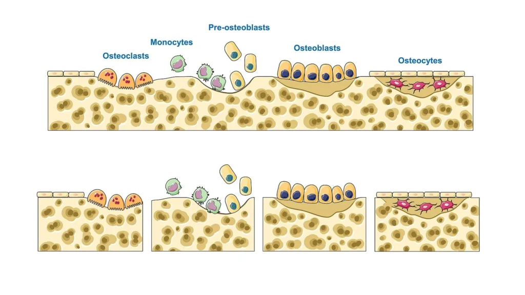
“Bone remodeling cycle” by SMART-Servier Medical Art (Laboratories Servier), via Wikimedia Commons Licensed under CC BY-SA3.0

How do osteocytes, bone-lining cells, and the RANK- RANKL- OPG signaling pathway coordinate the activation phase of bone remodeling to ensure targeted osteoclast recruitment and skeletal homeostasis?

Exam Question

2. Resorption Phase

During the resorption phase, activated osteoclasts adhere to the bone surface and establish a specialized sealed microenvironment known as the resorption lacuna (Howship’s lacuna). Within this compartment, osteoclasts generate a ruffled border to increase surface area for secretion.

They actively secrete hydrogen ions (via proton pumps) to acidify the environment, dissolving the inorganic hydroxyapatite matrix, while releasing proteolytic enzymes (e.g., cathepsin K) to degrade the organic collagen matrix. This coordinated process results in the formation of a resorption pit and removal of structurally compromised bone.

“Bone remodeling cycle” by SMART-Servier Medical Art (Laboratories Servier), via Wikimedia Commons Licensed under CC BY-SA3.0

By what cellular specializations and biochemical mechanisms do osteoclasts establish the resorption lacuna and achieve coordinated degradation of both mineral (hydroxyapatite) and organic (collagen) components of bone matrix?

Exam Question

3. Reversal Phase

The reversal phase represents a critical transitional stage between bone resorption and formation. Following osteoclast apoptosis or migration, mononuclear reversal cells colonize the resorption surface.

These cells remove residual debris, modify the microenvironment, and secrete signaling molecules that recruit and differentiate osteoblast precursors. This phase ensures proper coupling between resorption and formation, preventing structural discontinuity and enabling precise spatial coordination of new bone deposition.

“Bone remodeling cycle” by SMART-Servier Medical Art (Laboratories Servier), via Wikimedia Commons Licensed under CC BY-SA3.0

How do reversal cells mediate the transition from osteoclastic resorption to osteoblastic formation, and what mechanisms ensure effective coupling between these phases to maintain structural continuity of bone?

Exam Question

4. Formation Phase

During the formation phase, osteoblasts synthesize and deposit osteoid, the unmineralized organic matrix of bone composed primarily of type I collagen fibers and non-collagenous proteins.

Osteoid provides a structural scaffold for subsequent mineralization. Osteoblasts align along the resorption surface and coordinate matrix deposition in a highly regulated manner, restoring bone architecture. Over time, some osteoblasts differentiate into osteocytes, become bone-lining cells, or undergo apoptosis.


“Bone remodeling cycle” by SMART-Servier Medical Art (Laboratories Servier), via Wikimedia Commons Licensed under CC BY-SA3.0

How do osteoblasts coordinate osteoid synthesis, spatial organization, and differentiation into osteocytes or lining cells, and how does this process restore bone architecture following resorption?

Exam Question

5. Mineralization Phase

The mineralization phase involves the deposition of inorganic minerals within the osteoid matrix. Calcium and phosphate ions precipitate to form hydroxyapatite crystals, which integrate with collagen fibers to confer mechanical strength and rigidity.

This process occurs in two stages: an initial rapid phase followed by a slower maturation phase that may continue for months. Proper mineralization is essential for restoring the functional load-bearing capacity of bone.

“Bone remodeling cycle” by SMART-Servier Medical Art (Laboratories Servier), via Wikimedia Commons Licensed under CC BY-SA3.0

How does the process of osteoid mineralization proceed at the molecular level, including hydroxyapatite crystal formation, and how does this process restore the mechanical strength of bone over time?

Exam Question

SUMMARY TABLE

Scroll to Top