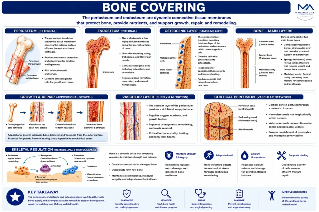
Bone Architecture
MYO CORE
Bone Covering
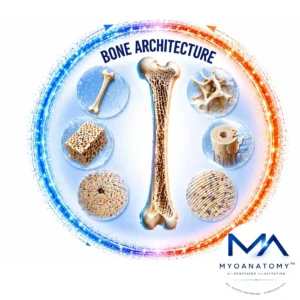
Bone coverings consist of specialized connective tissue membranes that envelop the external and internal surfaces of bone. The periosteum forms the highly vascular outer covering responsible for appositional growth, repair, and tendon attachment, whereas the endosteum lines internal osseous surfaces and regulates continuous bone remodeling through osteogenic cellular activity.
OVERVIEW
Bone covering represents the dynamic interface between skeletal tissue and its surrounding biological environment.
Externally, the periosteum provides mechanical protection, vascular supply, sensory innervation, and a reservoir of osteoprogenitor cells essential for growth and fracture healing.
Internally, the endosteum forms a thin osteogenic membrane lining the medullary cavity, trabecular surfaces, and Haversian systems, where it coordinates bone formation, resorption, and metabolic adaptation.
Together, these membranes maintain skeletal integrity by supporting bone growth, facilitating repair, regulating mineral homeostasis, and enabling lifelong structural remodeling in response to physiological and mechanical demands.
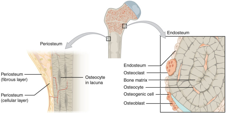
“Periosteum and Endosteum” – OpenStax College, Anatomy & Physiology, Connexions Web site via Wikimedia Commons. Licensed under CC BY 3.0.
ANATOMY
Periosteum
The periosteum is a dense connective tissue membranecovering the external surface of bone, except at articular cartilage–covered regions.
It consists of two distinct layers:
an outer fibrous layer, composed of collagen fibers, providing mechanical protection and structural support
an inner osteogenic layer, containing osteoprogenitor cells capable of differentiating into osteoblasts
The periosteum plays a critical role in bone growth (appositional growth), fracture repair, and serves as a site for attachment of tendons and ligaments via Sharpey’s fibers, enabling efficient force transmission between muscle and bone.
Exam Question
How does the bilaminar structure of the periosteum, including its fibrous and osteogenic layers, enable coordinated mechanical force transmission, appositional bone growth, and fracture repair, and what is the functional significance of Sharpey’s fibers in this context?
Endoosteum
The endosteum is a thin, highly cellular connective tissue lining the internal surfaces of bone, including the medullary cavity, trabecular surfaces, and Haversian canals.
It contains osteogenic cells, including osteoblasts and osteoclasts, and is actively involved in bone remodeling, regulating bone formation and resorption.
Functionally, the endosteum plays a central role in metabolic regulation of bone tissue, maintaining mineral homeostasis and enabling continuous structural adaptation to mechanical and physiological demands.
Exam Question
How does the cellular composition of the endosteum regulate the dynamic balance between bone formation and resorption, and how does this contribute to continuous structural adaptation and mineral homeostasis within the skeletal system?
Osteogenic Layer
The osteogenic layer (cambium layer) is the inner cellular component of the periosteum and serves as the principal biological center for skeletal growth, repair, and regeneration.
Rich in osteoprogenitor cells, vascular networks, and regulatory growth factors, it forms a highly active interface between the periosteum and cortical bone. Through continuous cellular renewal and differentiation, the osteogenic layer maintains bone homeostasis, supports structural adaptation to mechanical demands, and provides the regenerative potential required for lifelong skeletal maintenance.
Exam Question
How does the osteogenic layer function as the primary regenerative compartment of the periosteum, and how do its cellular and molecular components support skeletal growth, adaptation, repair, and long-term bone homeostasis?
Bone Forming Layer
The bone-forming function of the osteogenic layer is mediated by osteoprogenitor cells that differentiate into osteoblasts under the influence of signaling pathways such as BMPs, Runx2, Osterix, and Wnt/β-catenin.
These osteoblasts synthesize osteoid composed primarily of type I collagen and extracellular matrix proteins, which subsequently mineralize to form mature bone tissue.
This continuous process enables appositional growth, replacement of damaged bone, preservation of skeletal strength, and ongoing structural remodeling throughout life.
Exam Question
How do osteoprogenitor cells differentiate into osteoblasts, and how does osteoblast-mediated osteoid production and mineralization contribute to bone growth, remodeling, structural integrity, and skeletal regeneration?
Growth and Repair
The osteogenic layer is the primary site of appositional bone growth, where new bone is deposited on the external surface of existing cortical bone.
This process increases bone diameter and cortical thickness while preserving structural strength, enabling the skeleton to adapt continuously to developmental changes and mechanical loading throughout life.
The osteogenic layer plays a critical role in fracture healing and skeletal regeneration. Following injury, its cellular population rapidly proliferates and migrates toward the damaged region, contributing to callus formation, woven bone deposition, and subsequent remodeling into mature lamellar bone.
Through this regenerative capacity, the osteogenic layer supports restoration of skeletal continuity, structural integrity, and long-term functional adaptation.
Exam Question
How do osteoprogenitor cells differentiate into osteoblasts, and how does osteoblast-mediated osteoid production and mineralization contribute to bone growth, remodeling, structural integrity, and skeletal regeneration?
Vascular Layer
The vascular layer of the periosteum is a highly vascularized connective tissue compartment that forms the principal circulatory interface between the systemic bloodstream and skeletal tissue. Rich in arteries, veins, capillaries, lymphatics, and neurovascular structures, it supplies oxygen, nutrients, growth factors, and signaling molecules to the periosteum, cortical bone, and osteogenic layer.
Through its intimate association with bone-forming cells, this layer plays a central role in skeletal growth, metabolic maintenance, remodeling, and tissue viability while providing the biological support required for continuous adaptation of the skeleton throughout life.
Exam Question
How does the vascular layer of the periosteum function as the principal circulatory interface of bone, and how does its vascular network support skeletal growth, metabolic maintenance, tissue viability, remodeling, and lifelong adaptation of the skeleton?
Cortical Perfusion
The vascular layer contributes significantly to the blood supply of compact bone through numerous periosteal vessels that penetrate the cortex via Volkmann canals.
These vessels nourish the outer cortical regions, support osteocyte survival, maintain mineral homeostasis, and facilitate continuous bone remodeling.
By integrating with the Haversian vascular system, they establish an extensive microcirculatory network that preserves the structural and metabolic integrity of skeletal tissue.
Exam Question
How do periosteal vessels, Volkmann canals, and the Haversian vascular system cooperate to establish cortical perfusion, and how does this microcirculatory network maintain osteocyte survival, mineral homeostasis, and continuous bone remodeling?
Skeletal Regulation
Beyond its nutritive function, the vascular layer serves as a critical regulator of skeletal growth and regeneration.
Its vascular networks deliver osteogenic cells, cytokines, and growth factors that stimulate osteoblast activity and new bone formation.
Following injury, these vessels initiate angiogenesis, recruit reparative cells, and restore local perfusion, making revascularization one of the earliest and most essential events in fracture healing, callus formation, and successful bone regeneration.
Exam Question
How does the vascular layer regulate skeletal growth and regeneration through the delivery of osteogenic cells, cytokines, and growth factors, and why are angiogenesis and revascularization essential for fracture healing, callus formation, and successful bone repair?
SUMMARY TABLE