Skull Veins
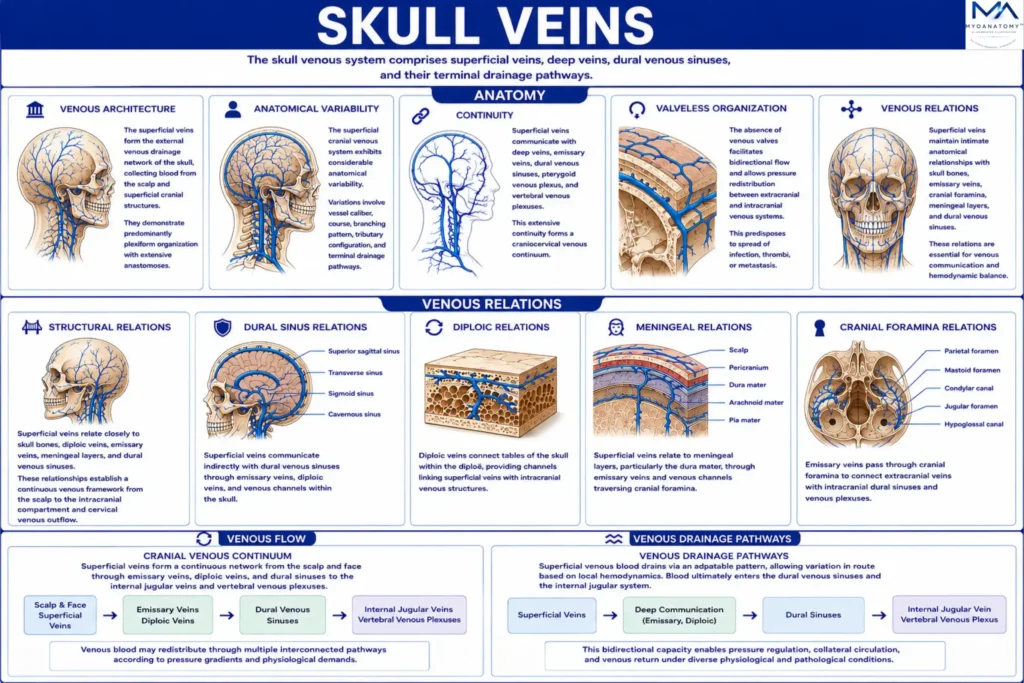
The skull venous system comprises superficial veins, deep veins, dural venous sinuses, and their terminal drainage pathways.

Superficial Veins
MYO CORE
Core Anatomy
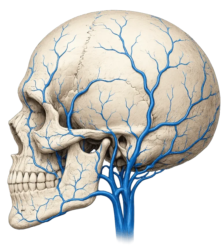
The superficial veins form the external venous drainage network of the skull, collecting blood from the scalp and superficial cranial structures. Extensive communications with deep veins, dural venous sinuses, emissary veins, and vertebral venous plexuses establish important connections between extracranial and intracranial venous pathways.
OVERVIEW
Unlike the arterial system, which follows a relatively predictable hierarchical branching pattern, the superficial venous system demonstrates marked anatomical variability, extensive anastomotic communications, and direct continuity with deeper cranial and intracranial venous pathways.
Rather than functioning as isolated vessels, the superficial veins represent the initial component of a continuous cranio-cervical venous network extending from the superficial tissues of the head to the dural venous sinuses, internal jugular veins, and vertebral venous plexuses.
Through numerous communicating channels, this system permits dynamic redistribution of blood flow according to changing physiological and hemodynamic demands, while simultaneously linking extracranial and intracranial venous compartments.
ANATOMY
Venous Architecture
A defining feature of the superficial cranial venous system is its predominantly plexiform organization. Numerous venous anastomoses connect adjacent drainage territories, producing an extensive network of communicating channels rather than a strictly hierarchical arrangement.
This architecture provides substantial hemodynamic redundancy. When individual vessels become compressed, narrowed, or obstructed, venous blood may be redirected through alternative superficial veins, deep cranial veins, emissary veins, diploic veins, vertebral venous plexuses, or cervical drainage pathways. Consequently, venous return from the craniofacial region depends upon an integrated network of parallel channels rather than a single dominant route.
This extensive interconnection is fundamental to the adaptability and resilience of cranial venous drainage.
Exam Question
Why is the plexiform and highly anastomotic organization of the superficial cranial venous system critical for maintaining venous return during vascular obstruction?
Anatomical Variability
The superficial cranial venous system exhibits considerably greater anatomical variability than the corresponding arterial circulation. Variations commonly involve vessel caliber, course, branching pattern, tributary configuration, anastomotic communications, and terminal drainage pathways.
Such variability reflects the developmental plasticity of the embryonic venous plexuses from which the cranial venous system originates. During development, selective enlargement, regression, and persistence of venous channels create individualized patterns of venous drainage while preserving overall functional efficiency.
Although specific venous configurations vary among individuals, the fundamental organization of cranial venous drainage remains consistent, ensuring effective venous return through multiple alternative pathways.
Exam Question
How does developmental remodeling of embryonic venous plexuses contribute to the marked anatomical variability observed in the superficial cranial venous system?
Extracranial-Intracranial Continuity
One of the most distinctive features of superficial cranial venous anatomy is its direct continuity with intracranial venous structures. Communication occurs through emissary veins, diploic veins, ophthalmic veins, pterygoid venous plexuses, and dural venous sinuses.
These channels establish direct anatomical and functional connections between superficial tissues of the scalp and face and the intracranial compartment. Consequently, the superficial venous system contributes not only to extracranial drainage but also to the overall regulation of cranial venous outflow.
From a neurovascular perspective, superficial veins function as extracranial extensions of the broader intracranial venous circulation, creating an integrated drainage network that links the scalp, skull, meninges, and brain.
Exam Question
Evaluate the anatomical and functional significance of direct continuity between superficial cranial veins and intracranial venous structures.
Valveless Organization
A fundamental principle of cranial venous anatomy is the relative absence of functional venous valves throughout much of the superficial and intracranial venous circulation. This characteristic extends to superficial cranial veins, emissary veins, diploic veins, ophthalmic veins, vertebral venous plexuses, and dural venous sinuses.
Unlike peripheral veins, where valves maintain predominantly unidirectional blood flow, cranial venous circulation is governed primarily by regional pressure gradients. This arrangement permits considerable flexibility within the system and facilitates adaptation to changes in posture, respiration, intracranial pressure, cerebral blood flow, and cervical venous pressure.
The valveless nature of these vessels is therefore a key structural feature underlying the unique physiology of cranial venous drainage.
Exam Question
How does the absence of functional venous valves influence pressure regulation and blood-flow dynamics within cranial venous circulation?
Bidirectional Flow
Because many cranial veins lack functional valves, blood flow may occur in either direction depending upon local hemodynamic conditions.
Under normal circumstances, venous blood follows pathways of least resistance toward the internal jugular veins and vertebral venous plexuses. However, alterations in intracranial pressure, thoracic pressure, respiratory mechanics, cervical venous pressure, or vascular obstruction may modify pressure gradients and reverse local flow patterns.
This bidirectional capability allows the cranial venous system to function as a dynamic pressure-regulating network capable of redistributing blood between superficial, deep, extracranial, and intracranial venous territories. Such adaptability is particularly evident within emissary veins, ophthalmic veins, diploic veins, and vertebral venous plexuses.
Exam Question
Why is bidirectional venous flow considered a fundamental adaptive mechanism of the cranial venous system?
Venous Relations
Structural Relations
The superficial cranial venous system maintains intimate anatomical relationships with the skull bones, diploic veins, emissary veins, cranial foramina, meningeal layers, and dural venous sinuses. These relationships establish a continuous venous framework extending from the scalp to the intracranial compartment and ultimately to the major cervical outflow pathways.
Through these structural connections, superficial veins participate not only in extracranial drainage but also in pressure redistribution, collateral venous circulation, and communication between superficial and deep cranial venous territories.
Exam Question
How do the structural relationships between superficial veins, skull bones, emissary veins, and dural sinuses facilitate collateral cranial venous circulation?
Dural Sinus Relations
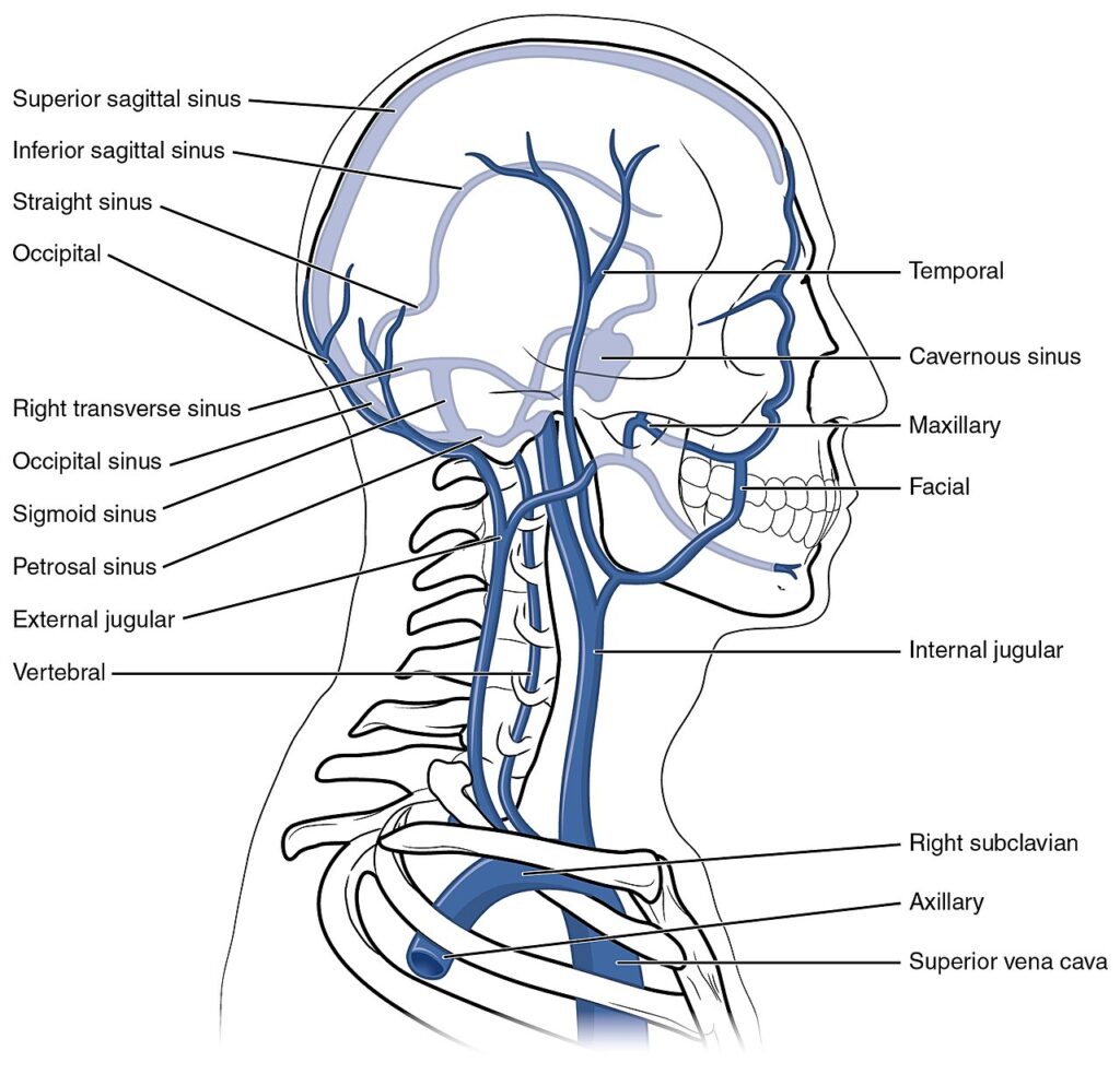
The superficial veins communicate indirectly with the dural venous sinuses through emissary veins, diploic veins, ophthalmic veins, and pterygoid venous plexuses. The dural sinuses constitute the principal intracranial collecting channels and ultimately receive blood originating from both cerebral and superficial cranial venous territories.
Consequently, the superficial venous system forms the extracranial component of a larger venous network that converges within the dural sinuses before draining through the internal jugular veins.
Exam Question
Explain how superficial cranial veins communicate with dural venous sinuses and why this relationship is essential for intracranial venous drainage.
Diploic Relations
Within the diploë of the cranial vault lies a network of valveless diploic veins situated between the outer and inner tables of the skull. These veins establish communication between superficial scalp veins and intracranial dural venous sinuses, creating an intermediate venous compartment within the cranial bones themselves.
Through these channels, venous blood may be redistributed between extracranial and intracranial systems according to changing pressure gradients, making the diploic veins an important anatomical bridge between superficial and deep cranial venous circulation.
Exam Question
What is the functional importance of diploic veins as intermediary channels linking superficial cranial veins with intracranial venous sinuses?
Meningeal Relations
The superficial venous system maintains an important relationship with the meningeal layers, particularly the dura mater. The dural venous sinuses are located between layers of dura and receive blood from both cerebral and extracranial sources through communicating venous channels.
As a result, superficial cranial veins are anatomically linked to meningeal venous structures despite remaining predominantly extracranial. This relationship forms part of the continuous venous pathway connecting superficial tissues with the intracranial compartment.
Exam Question
How do meningeal layers and dural venous sinuses integrate superficial cranial venous drainage into the intracranial venous system?
Cranial Foramina Relations
Communication between superficial and intracranial venous systems occurs largely through emissary veins traversing cranial foramina and osseous canals. Important pathways include the parietal foramen, mastoid foramen, condylar canal, and multiple skull-base openings transmitting emissary veins between extracranial veins and intracranial dural sinuses.
These foramina function as anatomical gateways through which venous blood may pass between superficial and intracranial compartments, contributing to collateral drainage, pressure equalization, and continuity of venous outflow.
Exam Question
Why are cranial foramina and emissary veins considered key anatomical gateways between extracranial and intracranial venous compartments?
Skull Bone Relations
The superficial veins are closely related to the cranial vault and overlying soft tissues. Many vessels course within the dense connective tissue layer of the scalp immediately superficial to the calvaria, while others communicate with diploic and emissary veins located within the skull itself.
Consequently, the cranial bones serve not only as protective structures but also as conduits for venous communication through diploic channels and emissary pathways. This relationship establishes direct continuity between superficial extracranial veins and intracranial venous structures.
Exam Question
Evaluate the role of the cranial vault as both a protective structure and a conduit for venous communication between superficial and intracranial territories.
Venous Flow
Cranial Venous Continuum
The superficial veins should not be viewed as isolated extracranial vessels but rather as the initial component of an integrated cranial venous continuum linking the scalp, skull, meninges, brain, and cervical venous system.
Superficial Veins → Emissary Veins → Diploic Veins → Dural Venous Sinuses → Internal Jugular Veins / Vertebral Venous Plexuses
Within this continuum, venous blood may be redistributed through multiple interconnected pathways according to regional pressure gradients and physiological requirements. This concept is fundamental to understanding both the anatomy and physiology of cranial venous drainage.
Exam Question
How does the concept of a cranial venous continuum improve understanding of venous communication between the scalp, skull, meninges, brain, and cervical drainage pathways?
Venous Drainage Pathways
The overall pattern of superficial cranial venous drainage follows a coordinated yet highly adaptable sequence. Blood originating within the scalp and facial tissues initially enters the superficial venous network before passing into deeper communicating channels, including emissary veins, diploic veins, ophthalmic veins, and deep cranial veins.
Subsequently, venous blood enters the dural venous sinuses, which function as the principal intracranial collecting reservoirs. From these sinuses, blood exits the cranial cavity primarily through the internal jugular veins and vertebral venous plexuses before returning to the systemic circulation.
Scalp & Face → Superficial Veins → Emissary Veins / Deep Cranial Veins → Dural Venous Sinuses → Internal Jugular Veins / Vertebral Venous Plexuses
Although this represents the predominant drainage pathway, the extensive anastomotic architecture of the cranial venous system allows substantial variation in the precise route taken by blood flow, ensuring effective venous return under both physiological and pathological conditions.
Exam Question
Trace the principal pathway of superficial cranial venous drainage and explain how extensive anastomoses permit alternative routes of venous return.
SUMMARY TABLE